which is the advantage of positron emission tomography (pet) over functional mri (fmri)?
As technology continues to advance, the medical field has seen significant developments in imaging techniques. Two widely used methods are Positron Emission Tomography (PET) and Functional Magnetic Resonance Imaging (fMRI). While both hold immense value in medical diagnosis and research, PET offers several advantages over fMRI. Understanding the advantages of PET can shed light on why it is often preferred in certain situations.
Understanding Positron Emission Tomography (PET)
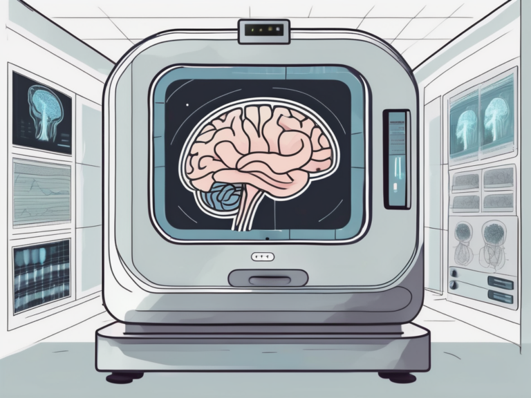
PET is an imaging technique that allows physicians and researchers to visualize the functioning of organs and tissues inside the human body. It relies on the detection of gamma rays emitted by radioactive substances known as radiotracers. By injecting these tracers into the body, PET scanners can create detailed images that indicate areas of high tracer uptake.
These images provide valuable insight into the metabolic and biochemical processes occurring within different organs and tissues. PET scans are commonly used in the fields of oncology, cardiology, neurology, and psychiatry.
The Science Behind PET
Understanding the science behind PET can help explain its advantages over fMRI. PET utilizes a radioactive tracer that emits positrons, which are particles with positive charge. When a positron collides with an electron, they annihilate each other, resulting in the emission of two gamma rays. These gamma rays are then detected by the PET scanner and used to create images of the internal structures.
Through this process, PET measures the distribution and concentration of radiotracers within the body. By injecting different radiotracers, specific organs or processes can be targeted, providing valuable information about their functioning.
How PET Scans Work
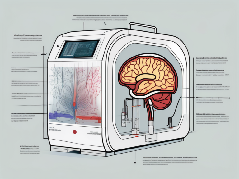
PET scanners consist of two main components: the scanner itself and a computer system. The patient is positioned inside the scanner, which consists of a large ring that contains multiple radiation detectors. These detectors measure the gamma rays emitted by the radiotracers in the body.
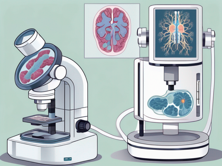
Once the scan is complete, the data is sent to a computer system that reconstructs the images and presents them to the healthcare provider or researcher. This allows for detailed analysis and interpretation of the PET scan results.
It is important to note that PET scans are non-invasive and generally safe, with minimal side effects. The radiotracers used in PET imaging have short half-lives, which means they decay quickly and are eliminated from the body through natural processes. This ensures that the radiation exposure to the patient is minimal.
Furthermore, PET scans can provide valuable information about the stage and progression of diseases, such as cancer. By visualizing the metabolic activity of tumors, PET scans can help determine the extent of cancer spread and guide treatment decisions.
In addition to diagnosing and monitoring diseases, PET scans also play a crucial role in research. They allow scientists to study various aspects of human physiology and pathology, aiding in the development of new treatments and therapies.
Delving into Functional MRI (fMRI)
fMRI is another imaging technique commonly used in the medical field. It measures changes in blood oxygenation levels in the brain to identify regions of neural activity. This non-invasive method provides functional information about the brain and has become an invaluable tool in neuroscience and clinical research.
The Basics of fMRI
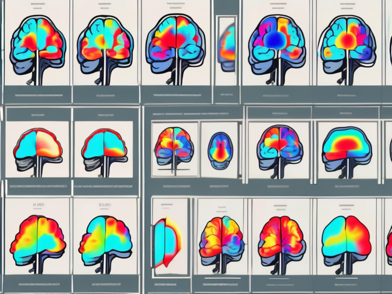
fMRI relies on the principle that increased neural activity leads to increased blood flow and oxygenation in the corresponding brain regions. By detecting changes in blood oxygenation levels, fMRI can identify brain areas that are involved in specific tasks or processes.
fMRI is often used to study brain disorders, cognitive processes, language processing, and emotional responses. It has greatly contributed to our understanding of the human brain.
The Process of fMRI Scanning
During an fMRI scan, the patient lies inside a magnetic resonance imaging (MRI) machine. The MRI machine generates a strong magnetic field that aligns the hydrogen atoms in the body. Radio frequency pulses are then used to perturb the alignment and measure the response of the hydrogen atoms.
This response is used to create detailed images of the brain, and changes in blood oxygenation levels are detected through specific sequences in the MRI scan. These changes are then associated with neural activity and provide insights into brain functioning.
One fascinating aspect of fMRI is its ability to capture real-time brain activity. Researchers can track changes in neural responses as participants engage in various tasks, allowing for a dynamic view of brain function. This capability has opened up new avenues of research in fields such as psychology, neurology, and even marketing.
Moreover, fMRI technology continues to evolve, with advancements in imaging resolution and data analysis techniques. These improvements enable researchers to delve deeper into the complexities of the brain, unraveling intricate networks of connectivity and shedding light on the mechanisms underlying behavior and cognition.
Comparing PET and fMRI
Similarities Between PET and fMRI
While PET and fMRI are distinct imaging techniques, they share some similarities. Both provide functional information about the human body and are non-invasive. They have had significant impacts on medical research and clinical practice, contributing to advancements in several fields.
Both PET and fMRI require a scanner and computer system to interpret the data obtained. Additionally, they both generate images that highlight areas of interest, helping healthcare providers and researchers make informed decisions.
Another key similarity between PET and fMRI is their ability to detect changes in the body’s physiology. PET scans use radiotracers to visualize metabolic processes, such as glucose metabolism, which can be crucial in diagnosing conditions like cancer or heart disease. On the other hand, fMRI measures changes in blood flow and oxygen levels in the brain, offering insights into neural activity and brain function.
Furthermore, both imaging techniques play a vital role in personalized medicine by providing detailed information about an individual’s unique physiology. This personalized approach allows for tailored treatment plans and improved patient outcomes.
Key Differences Between PET and fMRI
Despite their similarities, PET and fMRI have unique characteristics that set them apart. These differences are critical in understanding why PET offers advantages over fMRI in certain situations.
One significant difference is the type of information provided by each imaging method. PET offers insights into the metabolic and biochemical processes of various organs and tissues. In contrast, fMRI primarily focuses on brain activity and connectivity.
An additional difference lies in the temporal resolution of the imaging techniques. PET scans have a longer time frame, typically measuring minutes to hours, while fMRI provides near real-time information, with measurements made in seconds. This distinction impacts the types of processes and events that can be effectively studied using each method.
Moreover, PET imaging is particularly useful in oncology for detecting tumors and assessing treatment response. The ability of PET scans to visualize cellular function and metabolism aids in identifying cancerous tissues and monitoring their changes over time. In comparison, fMRI is widely used in neuroscience research to map brain activity and understand cognitive processes, such as memory formation and decision-making.
Advantages of PET over fMRI
Superior Imaging Capabilities of PET
PET scans provide highly detailed images, offering superior spatial resolution compared to fMRI. This allows for precise localization of areas of interest, making PET an optimal choice for identifying abnormalities and specific patterns of activity within the body.
The ability of PET to provide biochemical and metabolic information is another key advantage. By using radiotracers targeted to specific processes or organs, PET can offer crucial insights into diseases and conditions. This advantage is especially valuable in oncology, allowing for early detection, staging, and monitoring of various cancers.
Furthermore, PET imaging is not affected by the magnetic fields used in fMRI, making it a preferred choice for patients with metallic implants or devices that are contraindicated for fMRI scans. This makes PET a versatile option for a wider range of patients, ensuring that they can still receive accurate diagnostic imaging without concerns about compatibility issues.
PET’s Versatility in Medical Diagnosis
PET’s versatility makes it a valuable tool in medical diagnosis. It can be used to assess organ function, identify the presence and location of tumors, diagnose neurological disorders, and evaluate the efficacy of treatments. PET is also beneficial for patients with conditions that may require surgical interventions, as it provides detailed preoperative images, aiding in surgical planning.
Moreover, PET imaging can be combined with other imaging modalities such as CT or MRI to provide a more comprehensive evaluation of a patient’s condition. This multi-modality approach enhances diagnostic accuracy and allows for a more thorough assessment of complex medical cases. The integration of PET with other imaging techniques further highlights its adaptability and effectiveness in clinical practice.
Potential Limitations of PET and fMRI
Drawbacks of PET Scans
Like any medical imaging technique, PET scans have some limitations. The use of radiation and the need for radiotracers can pose potential risks to patients. However, rigorous safety protocols and guidelines are in place to minimize these risks.
Another drawback of PET is the limited availability of radiotracers for specific organs or processes. The development of novel radiotracers is an ongoing area of research, aiming to expand the scope and applicability of PET imaging.
Despite these limitations, PET scans remain a valuable tool in the field of medical imaging. They are particularly useful in oncology for detecting and monitoring tumors, as well as in neurology for studying brain function and neurodegenerative diseases.
Limitations of fMRI Scans
fMRI also has its limitations. It is highly sensitive to patient movement, making it challenging to obtain accurate and reliable images. Additionally, fMRI can be affected by artifacts, such as magnetic susceptibility and image distortion.
Furthermore, fMRI primarily provides indirect measurements of neural activity. Although it offers valuable insights into brain function, it may not fully capture all aspects of neural processes, potentially limiting its comprehensive understanding.
Despite these challenges, fMRI has revolutionized the field of cognitive neuroscience. It has enabled researchers to map brain activity in real-time, leading to significant advancements in understanding cognitive processes such as memory, attention, and decision-making.
The Future of PET and fMRI
Technological Advancements in PET
Advancements in PET technology continue to improve its capabilities and expand its potential applications. The development of new radiotracers and imaging techniques allows for more precise and targeted imaging. Researchers are also exploring hybrid imaging systems that combine PET with other modalities, such as CT or MRI, further enhancing diagnostic capabilities.
One exciting area of development in PET technology is the use of artificial intelligence (AI) algorithms to analyze PET images. These algorithms can help in automating the process of image interpretation, leading to faster and more accurate diagnoses. Additionally, researchers are investigating the use of novel radiotracers that can target specific biological processes at the molecular level, opening up new possibilities for personalized medicine.
Innovations in fMRI Technology
fMRI technology is also advancing rapidly. Recent developments focus on improving spatial and temporal resolution, minimizing artifacts, and enhancing the sensitivity of the technique. These innovations aim to overcome existing limitations and provide even more valuable information about brain function and connectivity.
Another area of research in fMRI technology is the exploration of real-time fMRI, which allows for the monitoring of brain activity as it happens. This capability has significant implications for neurofeedback training and the study of dynamic brain processes. Furthermore, advancements in machine learning algorithms applied to fMRI data are enabling researchers to extract complex patterns of brain activity that were previously inaccessible.
As technology continues to evolve, the future of PET and fMRI holds great promise for revolutionizing medical imaging and neuroscience. The combination of these imaging modalities with cutting-edge developments in AI and machine learning is poised to unlock new insights into the complexities of the human body and brain, ultimately leading to improved diagnostic accuracy and treatment outcomes.