how do fmri scans work
In the world of medical imaging, one method that has revolutionized our understanding of the human brain is functional Magnetic Resonance Imaging, commonly known as fMRI scans. These non-invasive scans have helped researchers and medical professionals explore the intricacies of the brain, mapping brain activity and aiding in the diagnosis and treatment of various neurological disorders. In this article, we will delve into the ins and outs of fMRI scans, from their basic principles to their limitations and challenges.
Understanding the Basics of fMRI Scans
What is an fMRI Scan?
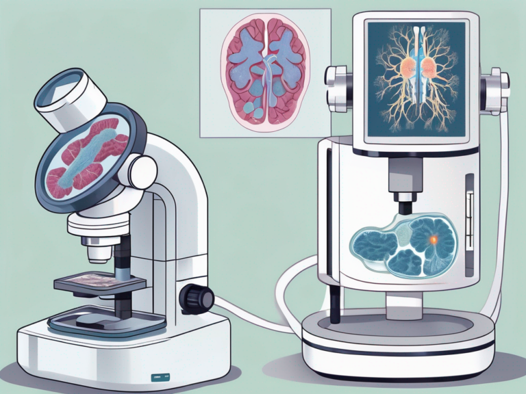
At its core, an fMRI scan is a technique that captures and maps brain activity by measuring changes in blood flow. This imaging method utilizes a powerful magnet and radio waves to generate detailed images of the brain, allowing researchers and medical professionals to observe brain regions that are active during certain tasks or are affected by neurological conditions.
During an fMRI scan, the individual lies inside a large, cylindrical machine called an MRI scanner. The scanner creates a magnetic field around the head, causing the hydrogen atoms in the body to align in a specific direction. When radio waves are sent through the body, these atoms emit signals that are picked up by the scanner and processed into images by a computer. By tracking the flow of oxygenated and deoxygenated blood in the brain, fMRI can pinpoint areas of increased neural activity.
The Importance of fMRI Scans in Medical Science
The significance of fMRI scans in medical science cannot be overstated. They provide valuable insights into brain functioning, offering a window into the complex processes that dictate our thoughts, emotions, and behaviors. By identifying abnormalities in brain activity, fMRI scans aid in the diagnosis and management of conditions such as epilepsy, stroke, and brain tumors.
Furthermore, fMRI technology has revolutionized the field of cognitive neuroscience by allowing researchers to explore the neural basis of various cognitive functions such as memory, attention, and decision-making. By studying how different regions of the brain communicate and interact during specific tasks, scientists can unravel the mysteries of the human mind and gain a deeper understanding of neurological disorders.
The Science Behind fMRI Scans
The Role of Magnetic Fields and Radio Waves
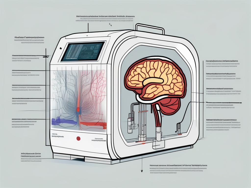
So, how do fMRI scans actually work? These scans rely on the interaction between magnetic fields and radio waves within the body. The patient lies inside a specially designed tube-shaped machine called an MRI scanner. This scanner generates a strong magnetic field that aligns the protons in water molecules within the body. Radio waves are then used to disturb these aligned protons, causing them to emit signals that are captured by the scanner.
The magnetic field produced by the MRI scanner is incredibly powerful, often measuring tens of thousands of times stronger than the Earth’s magnetic field. This strength is necessary to ensure that the protons align in a uniform manner, providing clear and accurate imaging results. The radio waves used in fMRI scans are carefully controlled in terms of frequency and intensity to target specific tissues and create detailed images of the brain.
How fMRI Scans Detect Brain Activity
By measuring blood flow in the brain, fMRI scans indirectly detect brain activity. When a particular brain region is engaged in a cognitive or motor task, it requires more oxygen and glucose. Consequently, blood flow to that region increases to meet the heightened demand. This increase in blood flow brings with it more oxygenated hemoglobin, which alters the magnetic properties of the brain tissue. The fMRI scanner picks up these changes, creating detailed images that highlight the active brain regions.
It’s fascinating to consider that fMRI technology has revolutionized our understanding of the human brain and its functions. The ability to visualize brain activity in real-time has opened up new avenues of research in fields such as neuroscience, psychology, and even artificial intelligence. Researchers can now observe how different areas of the brain communicate and coordinate during various tasks, providing valuable insights into cognition, behavior, and neurological disorders.
The Process of an fMRI Scan
Preparing for an fMRI Scan
Prior to an fMRI scan, certain preparations need to be made to ensure accurate results. Patients are typically advised to remove any metallic objects, such as jewelry and hairpins, as these can interfere with the magnetic field. It is crucial to follow these guidelines meticulously to guarantee the quality of the imaging results. Moreover, patients are usually instructed to refrain from consuming caffeine or any stimulants before the scan, as these substances can affect brain activity and alter the scan findings. This attention to detail in the pre-scan instructions helps in obtaining precise and reliable data.
Furthermore, individuals with claustrophobia may benefit from discussing their concerns with the healthcare provider, who may offer strategies to minimize discomfort during the scan. Techniques such as practicing relaxation exercises or using a mirror system that allows patients to see outside the scanner can be employed to alleviate anxiety. By addressing these issues beforehand, healthcare professionals can ensure a smoother and more successful scanning experience for all patients.
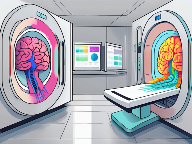
What to Expect During the Scan
During the fMRI scan, the patient lies still on a comfortable bed that slides into the scanner. The room is typically dimly lit to create a calming environment for the patient. Soft music or earplugs may be provided to reduce the noise generated by the MRI machine, which can sound like loud knocking or tapping. Patients are encouraged to communicate any discomfort or concerns to the MRI technologist, who is trained to assist and address any issues promptly.
It is crucial to remain as motionless as possible during the duration of the scan to avoid blurring the images. Even slight movements can distort the results, impacting the diagnostic quality of the scan. The MRI technologist will communicate with the patient through an intercom system and provide instructions for specific tasks or rest periods. These instructions are essential for certain types of fMRI scans that require the patient to perform cognitive tasks or respond to stimuli. The entire process is painless and generally takes around 30 to 60 minutes, depending on the specific requirements of the scan.
Interpreting fMRI Scan Results
Reading fMRI Images
Interpreting functional magnetic resonance imaging (fMRI) images is a complex process that requires a high level of expertise and a deep understanding of brain anatomy and function. These specialized scans provide valuable insights into brain activity by measuring changes in blood flow. Radiologists, neuroscientists, and other professionals carefully analyze the fMRI images to identify specific brain regions that are activated during different tasks or behaviors. By pinpointing these activation patterns, researchers can gain a better understanding of how the brain processes information and responds to various stimuli.
Furthermore, fMRI technology has revolutionized the field of neuroscience by allowing researchers to non-invasively study the brain’s inner workings in real-time. This imaging technique has opened up new possibilities for investigating cognitive processes, emotional responses, and neurological disorders. The data obtained from fMRI scans can help scientists map out neural networks, track changes in brain activity over time, and explore the underlying mechanisms of complex brain functions.
The Role of Radiologists in fMRI Analysis
Radiologists play a crucial role in the analysis of fMRI scans due to their specialized training in medical imaging interpretation. These experts are trained to recognize subtle differences in brain activity and to differentiate between normal and abnormal findings. By working closely with neurologists, neurosurgeons, and other healthcare professionals, radiologists help ensure accurate diagnoses and effective treatment plans for patients with neurological conditions.
Moreover, the collaboration between radiologists and researchers is essential for advancing our understanding of the human brain. By combining their clinical expertise with scientific knowledge, these professionals can contribute to groundbreaking discoveries in neuroscience and pave the way for innovative treatments for brain-related disorders. Radiologists bring a unique perspective to fMRI analysis, offering valuable insights that can shape the future of both clinical practice and scientific research.
The Limitations and Challenges of fMRI Scans
Potential Risks and Side Effects
While fMRI scans are generally safe, there are a few considerations to keep in mind. The strong magnetic fields generated during the scan can cause certain metallic objects, such as pacemakers or metal implants, to shift or heat up, posing potential risks to patients. Additionally, the confined space of the scanner may trigger anxiety or claustrophobia in some individuals. However, healthcare providers take necessary precautions to ensure patient safety and comfort.
One potential risk associated with fMRI scans is the interaction between the magnetic fields and metallic objects within the body. Pacemakers, for example, can be affected by the strong magnetic fields, potentially causing them to malfunction. This is why it is crucial for patients to inform their healthcare providers about any metal implants or devices they have before undergoing an fMRI scan. By doing so, healthcare professionals can take appropriate measures to minimize any potential risks and ensure the safety of the patient.
In addition to the physical risks, the confined space of the fMRI scanner can also be challenging for some individuals. The narrow tube-like structure of the scanner can trigger feelings of anxiety or claustrophobia, making it uncomfortable for certain patients. To address this, healthcare providers often offer strategies to help patients manage their anxiety, such as providing them with headphones to listen to music or offering relaxation techniques before the scan. By prioritizing patient comfort, healthcare professionals strive to create a positive experience during fMRI scans.
The Ongoing Debate About fMRI Accuracy
It is important to acknowledge that fMRI scans have their limitations. The accuracy of fMRI results has been a subject of ongoing debate within the scientific community. Like any diagnostic tool, fMRI scans have certain uncertainties and can yield false positives or false negatives. Researchers continue to refine the technology and explore alternative methods of fMRI analysis to enhance its reliability and clinical utility.
One of the main challenges in fMRI research is distinguishing between neural activity and noise. The brain is a complex organ, and fMRI scans capture a vast amount of data. Analyzing this data accurately requires sophisticated algorithms and statistical techniques. Researchers are constantly working on improving these methods to ensure that the fMRI results are as reliable and accurate as possible.
Another aspect of the ongoing debate about fMRI accuracy is the reproducibility of results. Reproducibility refers to the ability to obtain consistent results when the same experiment is repeated. Some studies have raised concerns about the reproducibility of fMRI findings, highlighting the need for rigorous experimental design and standardized protocols. Efforts are being made to address these concerns by promoting transparency in research practices and encouraging collaboration among scientists to validate and replicate fMRI studies.
In conclusion, fMRI scans have revolutionized the field of neuroscience, enabling researchers and medical professionals to delve into the intricate workings of the human brain. By understanding the basic principles of fMRI scans and the science behind them, we can appreciate their significance in medical science and their potential to transform our understanding of neurological disorders. However, it is important to recognize their limitations and ongoing challenges, urging continuous research and improvement to enhance the accuracy and reliability of fMRI scans.