what is an fmri
Functional magnetic resonance imaging (fMRI) is a powerful tool that has revolutionized our understanding of the human brain and its functioning. In this article, we will explore the basics of fMRI, its role in neuroscience, the process of an fMRI scan, interpreting fMRI results, its impact on medical diagnosis, and the ethical considerations surrounding its use.
Understanding the Basics of fMRI
Definition and Purpose of fMRI
fMRI, short for functional Magnetic Resonance Imaging, is a non-invasive neuroimaging technique that has revolutionized the field of neuroscience. It measures changes in blood flow in response to neural activity, providing valuable insights into how the brain functions. By detecting these changes, fMRI allows researchers and clinicians to map brain regions that are involved in various cognitive processes, such as memory, attention, and emotion regulation. This mapping of brain activity is crucial for understanding the complexities of the human brain and its intricate networks.
One of the primary purposes of fMRI is to investigate brain function and connectivity. By observing which areas of the brain are activated during specific tasks or stimuli, researchers can gain a deeper understanding of how different regions work together to produce thoughts, emotions, and behaviors. This information is not only essential for advancing our knowledge of the brain but also has practical applications in diagnosing and treating neurological and psychiatric disorders.
The Science Behind fMRI
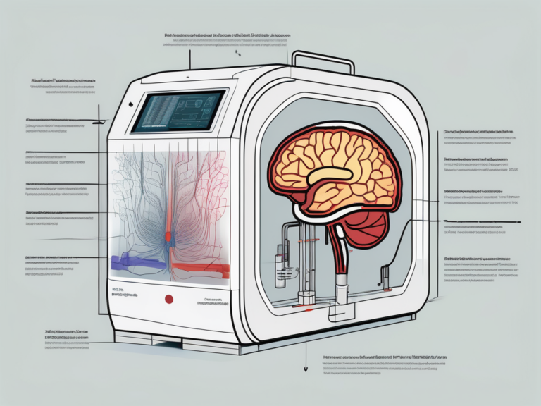
At the core of fMRI lies a fascinating scientific principle. When a specific region of the brain is engaged in a cognitive task, it requires more oxygenated blood supply to meet the increased metabolic demands of neural activity. This heightened demand for oxygen results in an influx of blood to the activated brain region, leading to an increase in blood flow. It is this hemodynamic response that forms the basis of fMRI imaging.
The fMRI machine detects these changes in blood flow by measuring the magnetic properties of the blood. Oxygen-rich blood has different magnetic properties than oxygen-poor blood, allowing the fMRI scanner to track the flow of blood to active brain regions with remarkable precision. The data collected from these magnetic signals is then processed using sophisticated algorithms to create detailed images of brain activity, known as activation maps. These maps provide researchers with a visual representation of which areas of the brain are involved in specific tasks, offering valuable insights into the neural mechanisms underlying various cognitive functions.
The Role of fMRI in Neuroscience
Brain Mapping and fMRI
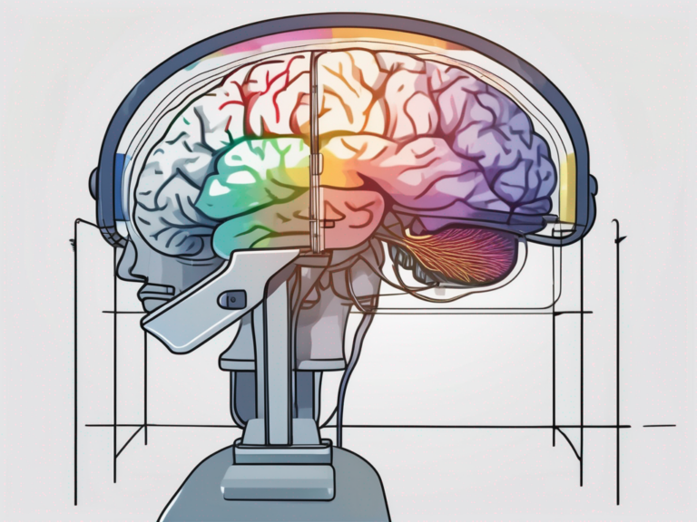
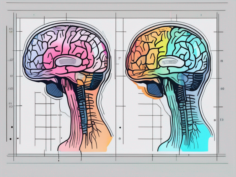
One of the major applications of fMRI is in brain mapping, which involves identifying the specific brain regions involved in various cognitive processes. By studying brain activity during specific tasks, researchers can create detailed maps that highlight the functional organization of the brain. This knowledge can help shed light on how different regions of the brain contribute to our perception, memory, language, and emotions.
Furthermore, fMRI technology has revolutionized the field of neuroscience by providing researchers with a non-invasive tool to visualize and understand the complex workings of the human brain. Through the use of advanced imaging techniques, scientists can now track changes in blood flow and oxygen levels in the brain, allowing them to pinpoint which areas are active during specific mental tasks.
fMRI in Cognitive Studies
fMRI also plays a crucial role in cognitive studies, allowing researchers to investigate the neural mechanisms underlying different cognitive processes. By comparing brain activity during different tasks or in different populations, researchers can gain insights into how our brains process information, make decisions, and adapt to changes.
Moreover, fMRI has opened up new avenues for studying brain disorders such as Alzheimer’s disease, schizophrenia, and autism. By analyzing the differences in brain activity patterns between healthy individuals and those with neurological conditions, researchers can develop a better understanding of the underlying causes of these disorders and explore potential treatment options.
The Process of an fMRI Scan
Preparing for an fMRI Scan
Prior to an fMRI scan, certain preparations are necessary. Patients may be asked to avoid caffeine or certain medications that could alter brain activity. Additionally, participants may undergo screening to ensure they do not have any contraindications for the scan, such as metallic implants that could interfere with the magnetic field. It is crucial for individuals to follow these guidelines to ensure the accuracy and safety of the imaging procedure. Furthermore, some research studies may require participants to complete questionnaires or cognitive assessments before the scan to gather additional information.
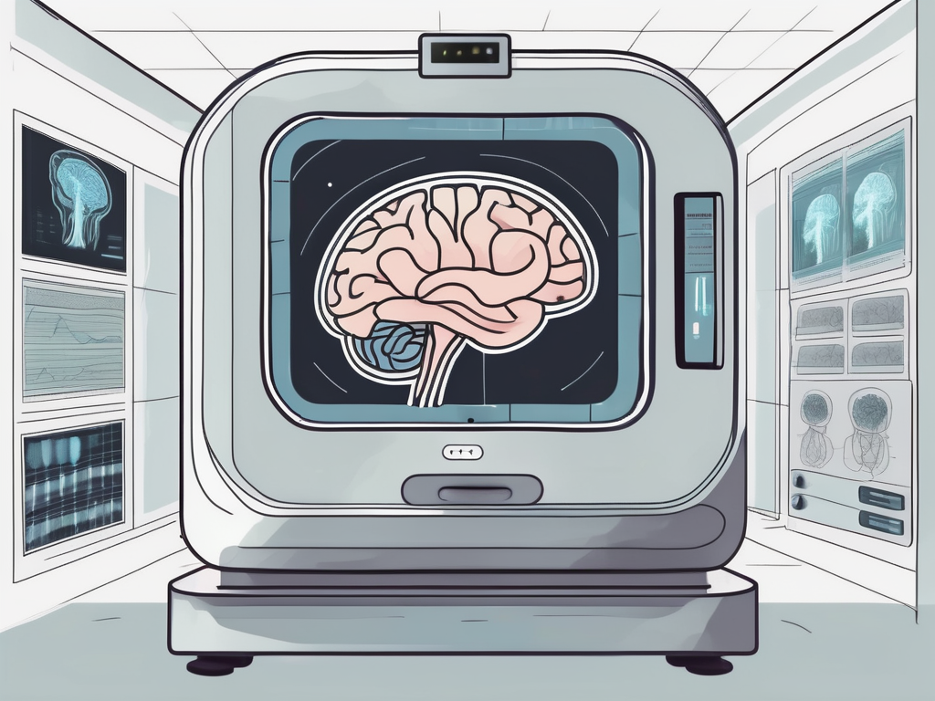
What Happens During an fMRI Scan
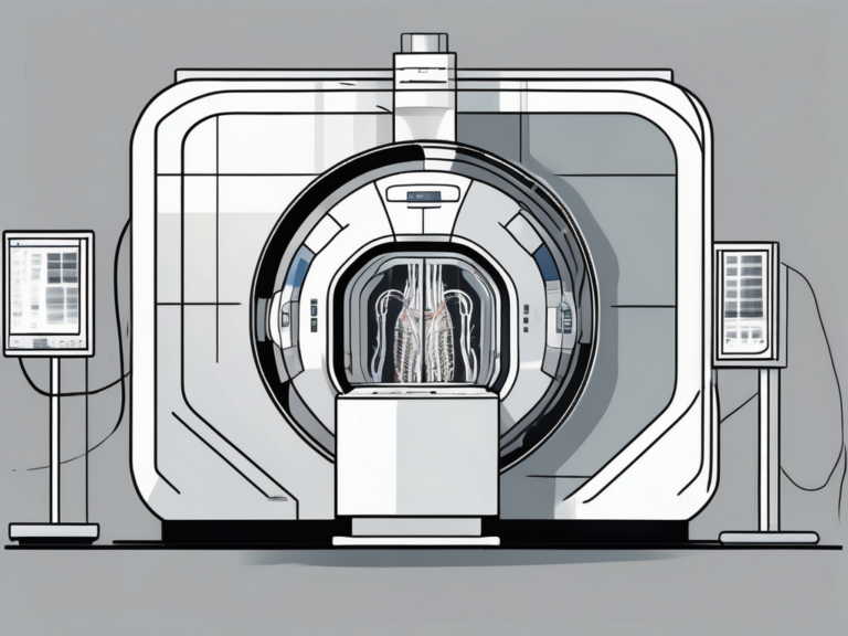
During an fMRI scan, the participant lies in a cylindrical tube that houses the magnet. The scan itself is painless, but the machine produces loud noises. To minimize discomfort, participants are often provided with earplugs or headphones. They are then instructed to perform specific cognitive tasks or rest quietly while the scan is taking place. The entire process is closely monitored by trained professionals. The fMRI machine uses a strong magnetic field and radio waves to create detailed images of the brain’s activity. As the participant engages in different tasks, changes in blood flow and oxygen levels in the brain are detected, providing valuable insights into brain function.
Moreover, fMRI scans have revolutionized the field of neuroscience by allowing researchers to observe the brain in action non-invasively. This technology has been instrumental in studying various cognitive processes such as memory, attention, and emotion. The data obtained from fMRI scans can help neuroscientists and clinicians better understand neurological disorders, track the effects of treatments, and even map out the intricate networks within the brain. By combining fMRI with other imaging techniques, such as structural MRI and diffusion tensor imaging, researchers can paint a comprehensive picture of brain structure and function.
Interpreting fMRI Results
Understanding fMRI Images
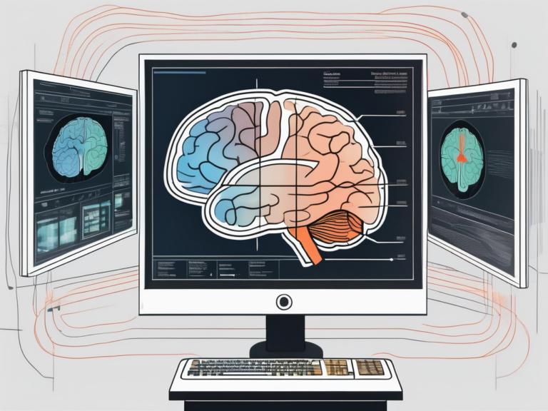
Interpreting fMRI images requires both expertise and experience. These images typically display brain activity as colorful overlays on anatomical brain scans. Researchers use statistical analysis to identify regions of the brain that show significant activity during a given task. However, it is important to note that fMRI measures blood flow, not neural activity directly, and thus, caution must be exercised when interpreting the results. Collaborative analysis between neuroscientists, statisticians, and imaging experts is crucial to ensure accurate interpretation.
Furthermore, the field of fMRI research is constantly evolving, with new methods and technologies being developed to improve the accuracy and reliability of results. For example, advancements in machine learning algorithms have enabled researchers to better distinguish true brain activity from noise in fMRI data. Additionally, multi-modal imaging approaches, combining fMRI with techniques such as EEG or MEG, offer a more comprehensive understanding of brain function by capturing different aspects of neural activity.
The Limitations and Challenges of fMRI Interpretation
While fMRI provides valuable insights into brain activity, there are inherent limitations and challenges associated with its interpretation. Factors such as head movement, individual differences, and the need for sophisticated analysis techniques can introduce variability and complexity. Additionally, fMRI is not suited for studying certain brain regions that are sensitive to movement artifacts, such as the brainstem. Therefore, a comprehensive understanding of both the strengths and limitations of fMRI is necessary for accurate interpretation.
To address some of these challenges, researchers are exploring innovative solutions such as real-time fMRI neurofeedback, which allows individuals to observe their own brain activity and learn to modulate it through techniques like meditation or cognitive training. This approach not only has therapeutic potential for conditions like anxiety and depression but also sheds light on the dynamic nature of brain function and its interaction with behavior. Despite the challenges, ongoing advancements in fMRI methodology continue to enhance our understanding of the human brain and its complexities.
The Impact of fMRI on Medical Diagnosis
fMRI in Neurological Disorders
fMRI has shown great promise in assisting in the diagnosis and treatment of various neurological disorders. By mapping brain activity, it can help identify aberrant patterns in individuals with conditions such as epilepsy, brain tumors, or neurodegenerative diseases. This information aids in surgical planning, therapeutic interventions, and monitoring treatment efficacy.
Furthermore, fMRI is also being increasingly utilized in the study of psychiatric disorders such as schizophrenia, depression, and anxiety. Researchers are exploring how fMRI can reveal neural correlates of these conditions, leading to more targeted and effective treatment strategies. The ability of fMRI to visualize brain activity in real-time provides valuable insights into the underlying mechanisms of these complex disorders.
Future Prospects of fMRI in Medicine
The field of fMRI in medicine continues to evolve rapidly, with ongoing research focusing on refining techniques, improving spatial and temporal resolution, and advancing our understanding of brain networks. This opens up exciting possibilities for the use of fMRI in personalized medicine, early disease detection, and targeted interventions.
Moreover, the integration of artificial intelligence and machine learning algorithms with fMRI data holds immense potential for revolutionizing medical diagnosis and treatment. These technologies can analyze vast amounts of fMRI data to identify subtle patterns and biomarkers that may not be apparent to the human eye. By leveraging these computational tools, healthcare professionals can make more accurate and timely clinical decisions, ultimately improving patient outcomes.
Ethical Considerations in fMRI Use
Privacy and Consent in fMRI Scans
As fMRI involves the collection of sensitive brain data, ensuring privacy and obtaining informed consent from participants is crucial. Researchers must follow strict ethical guidelines and take appropriate measures to protect the confidentiality of the data. Informed consent ensures that individuals understand the risks and benefits associated with participating in fMRI studies.
Moreover, privacy concerns extend beyond the data itself to the potential implications of the findings. Researchers must consider how the results of fMRI scans may impact an individual’s personal life, employment opportunities, or even insurability. Safeguarding the privacy of participants involves not only securing the data but also carefully considering the broader consequences of the research.
The Debate Over fMRI in Legal Cases
There is an ongoing debate regarding the use of fMRI in legal cases, particularly in assessing an individual’s truthfulness or detecting deception. While fMRI has the potential to provide valuable insights, its reliability and validity as a lie detector remain a topic of discussion among experts and within the legal community.
Furthermore, the use of fMRI in legal settings raises questions about the admissibility of neuroimaging evidence in court. Judges and legal scholars grapple with the challenge of balancing the potential benefits of fMRI technology in uncovering neural correlates of behavior with concerns about its interpretational limitations and the risk of undue influence on juries.
In conclusion, fMRI is a powerful tool that allows researchers and clinicians to investigate brain function, map brain regions involved in cognitive processes, and aid in the diagnosis and treatment of neurological disorders. However, its interpretation requires expertise and careful consideration of limitations, and ethical considerations must be addressed in its use. With ongoing advancements in technology and our understanding of the brain, fMRI holds great promise for further advancements in neuroscience and medicine.