eeg, meg, pet, and fmri are devices for measuring what?
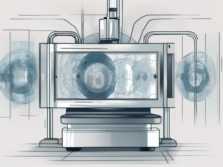
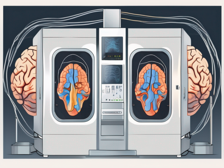
Brain imaging techniques have revolutionized the field of neuroscience, allowing experts to observe the inner workings of the human brain. Among the most widely used devices are electroencephalograms (EEG), magnetoencephalography (MEG), positron emission tomography (PET), and functional magnetic resonance imaging (fMRI). Each technique offers unique insights into the brain’s structure and function, making them invaluable tools in both clinical and research settings.
Understanding the Basics of Brain Imaging Techniques
The Science Behind Brain Imaging
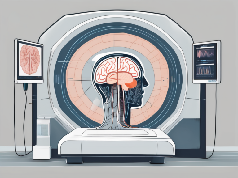
Before delving into the specifics of each brain imaging technique, it is crucial to grasp the basic principles underlying these technologies. At their core, these devices capture different physiological and anatomical processes to create detailed images of the brain. By visualizing brain activity, scientists can gain valuable information about cognition, emotion, and various neurological disorders.
One key aspect of brain imaging is its ability to map neural pathways and connectivity within the brain. This intricate network of connections is essential for understanding how different regions of the brain communicate and work together to perform various functions. Through advanced imaging techniques such as diffusion tensor imaging (DTI) and functional magnetic resonance imaging (fMRI), researchers can track the flow of information between brain regions in real-time, providing insights into complex cognitive processes.
The Role of Brain Imaging in Medical Diagnosis
Brain imaging techniques play a vital role in diagnosing and understanding neurological disorders. They provide crucial information to physicians, helping them identify abnormalities, locate lesions, and evaluate treatment effectiveness. Additionally, brain imaging aids in the early detection of conditions such as Alzheimer’s disease, epilepsy, and brain tumors, leading to improved patient outcomes.
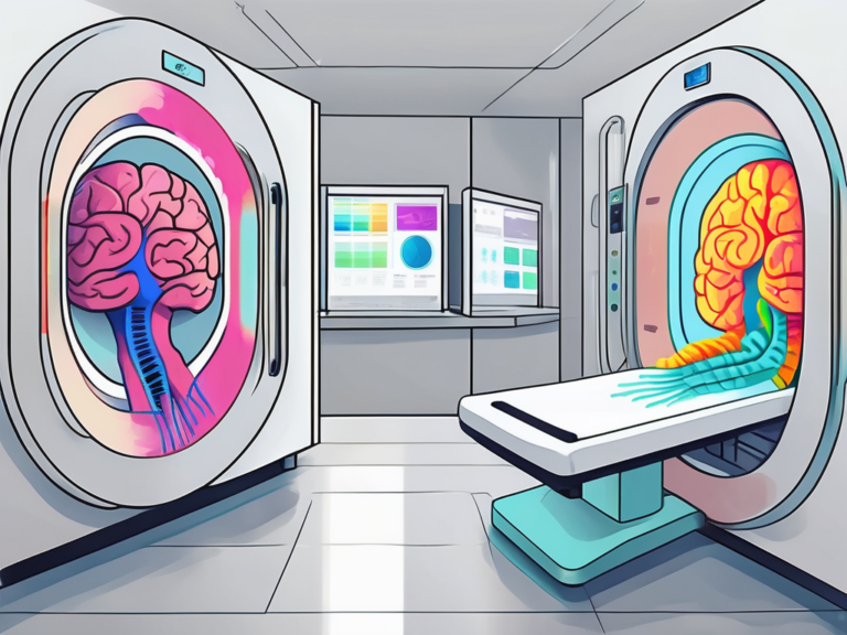
Moreover, brain imaging is instrumental in guiding surgical interventions for conditions that require precise localization, such as brain tumors and epilepsy surgery. By providing detailed maps of brain structures and functions, imaging techniques like magnetic resonance imaging (MRI) and positron emission tomography (PET) enable surgeons to plan and execute procedures with greater accuracy and safety. This targeted approach minimizes the risk of damage to healthy brain tissue and improves post-operative recovery for patients.
Exploring Electroencephalogram (EEG)
Electroencephalogram (EEG) is a non-invasive technique that measures the electrical activity of the brain by placing electrodes on the scalp. These electrodes detect the electrical impulses generated by neurons, allowing for the recording of brain waves. The data collected through EEG provides valuable insights into brain function and can help in understanding various cognitive processes.
One fascinating aspect of EEG is its ability to capture the brain’s electrical signals in real-time, offering a dynamic view of neural activity. Researchers and healthcare professionals utilize EEG to monitor brain health, study brain disorders, and even explore the intricacies of human consciousness.
How EEG Works
EEG measures the electrical activity of the brain, with electrodes placed on the scalp. By recording the brain’s electrical signals, experts can analyze and interpret neural patterns. The resulting readouts, called EEG graphs, display characteristic waveforms that correspond to different states of consciousness, such as sleep, wakefulness, and various cognitive processes.
Moreover, EEG technology has advanced significantly over the years, allowing for the development of portable EEG devices that can be used for continuous monitoring outside of clinical settings. This innovation has opened up new possibilities for studying brain activity in natural environments and everyday situations, providing researchers with a more comprehensive understanding of brain function.
Applications of EEG in Healthcare
EEG has proven instrumental in diagnosing and managing various neurological disorders. It is commonly used in epilepsy monitoring, helping physicians identify abnormal brain activity and determine appropriate treatment options. Furthermore, EEG enables the study of brain responses to external stimuli, facilitating research in areas like cognitive neuroscience, sleep disorders, and psychiatric conditions.
Additionally, EEG plays a crucial role in brain-computer interface (BCI) systems, where brain signals are used to control external devices or communicate with individuals who have limited motor functions. This groundbreaking application of EEG technology has the potential to enhance the quality of life for people with disabilities and revolutionize the field of neurotechnology.
Magnetoencephalography (MEG) Uncovered
The Functioning of MEG
MEG measures the magnetic fields generated by electrical currents within the brain, using highly sensitive sensors known as magnetoencephalometers. These sensors detect the tiny magnetic fields produced by neuronal activity, allowing researchers to analyze brain function with exceptional precision. MEG provides a temporal resolution unmatched by other techniques, enabling the investigation of rapid neural processes.
Furthermore, the non-invasive nature of MEG makes it a valuable tool in studying brain function in both healthy individuals and patients with neurological disorders. The data collected through MEG can be integrated with structural imaging techniques like MRI to create comprehensive maps of brain activity and connectivity, offering a holistic view of brain function.
The Use of MEG in Neurological Studies
MEG has proven particularly useful in studying brain dynamics and connectivity. Its ability to capture both cortical and deep brain activity helps researchers gain insights into various neurological conditions, such as stroke, epilepsy, and movement disorders. By mapping brain activity in real-time, MEG contributes to a deeper understanding of the brain’s complex network and related disorders.
Moreover, MEG is instrumental in pre-surgical planning for patients with epilepsy, as it helps identify the precise location of abnormal brain activity that may be causing seizures. This information is crucial for neurosurgeons to determine the most effective surgical approach, leading to better outcomes for patients with drug-resistant epilepsy.
Positron Emission Tomography (PET) Explained
Positron Emission Tomography (PET) is a powerful imaging technique that provides valuable insights into the metabolic processes of the human body. By utilizing the unique properties of radioactive tracers, PET allows for the visualization of physiological functions at a molecular level. This non-invasive procedure has revolutionized the field of medical imaging, particularly in the realm of neurology and oncology.
The Working Principle of PET
PET involves the injection of a radioactive tracer into the bloodstream, which emits positrons. As the tracer decays, it interacts with nearby electrons, producing gamma rays. These gamma rays are then detected by the PET scanner, generating three-dimensional images of tracer distribution within the brain. By visualizing metabolic processes, PET provides valuable information about brain function.
Furthermore, the data obtained from PET scans can be quantitatively analyzed to assess the rate of metabolic activity in different regions of the brain. This information is crucial for understanding the underlying mechanisms of various neurological disorders and can aid in the development of targeted treatment strategies.
PET in Detecting Metabolic or Physiological Disorders
PET has found widespread use in the diagnosis and management of various conditions, including cancer, heart disease, and neuroscience research. In neurology, PET is employed to detect metabolic abnormalities associated with conditions like Alzheimer’s disease, Parkinson’s disease, and epilepsy. It assists clinicians in determining the presence and extent of disease, guiding treatment protocols and monitoring therapeutic responses.
Moreover, PET imaging plays a vital role in oncology by enabling the early detection of tumors and metastases, as well as assessing treatment efficacy. The ability of PET to visualize metabolic changes in cancer cells has significantly improved the accuracy of cancer staging and personalized treatment planning.
Functional Magnetic Resonance Imaging (fMRI) Demystified
The Mechanism of fMRI
fMRI harnesses the power of magnetic resonance imaging (MRI) to measure blood flow changes in the brain, indirectly indicating neural activity. When a specific area of the brain becomes more active, blood flow to that region increases. By detecting these changes, fMRI produces detailed images that highlight brain activity patterns with remarkable precision.
Furthermore, the concept of fMRI is based on the principle that oxygenated and deoxygenated blood have different magnetic properties. This forms the basis of the blood-oxygen-level-dependent (BOLD) signal, which is the key measurement in fMRI. The BOLD signal allows researchers to infer neural activity by observing changes in blood oxygenation levels in different brain areas.
The Role of fMRI in Brain Function Analysis
fMRI has revolutionized our understanding of brain function. Through its non-invasive nature and high spatial resolution, fMRI has become a preferred technique for studying various cognitive processes, such as language, attention, and memory. It also aids in mapping brain regions responsible for specific functions, guiding surgical interventions, and improving therapeutic outcomes.
Moreover, fMRI is not limited to studying healthy brains. It has proven to be a valuable tool in investigating neurological and psychiatric disorders. Researchers use fMRI to compare brain activity patterns between individuals with different conditions, shedding light on the underlying neural mechanisms of diseases such as Alzheimer’s, schizophrenia, and depression. This insight is crucial for developing targeted treatments and interventions tailored to each patient’s unique brain activity profile.
Comparing EEG, MEG, PET, and fMRI
Similarities and Differences
While all four techniques provide valuable insights into the human brain, they differ significantly in terms of resolution, invasiveness, and the aspects of brain activity they capture. EEG and MEG offer exceptional temporal resolution but limited spatial resolution. On the other hand, PET and fMRI provide excellent spatial resolution but cannot capture rapid brain dynamics. Researchers often combine these techniques to obtain a comprehensive understanding of brain function and structure.
Electroencephalography (EEG) measures electrical activity in the brain by placing electrodes on the scalp. It is non-invasive and provides real-time information about brain function, making it ideal for studying brain waves during tasks such as sleep, attention, and memory. Magnetoencephalography (MEG) detects magnetic fields generated by neuronal activity and offers similar temporal resolution to EEG. MEG is particularly useful for localizing brain activity with high precision, aiding in the mapping of neural networks involved in various cognitive processes.
Choosing the Right Brain Imaging Technique
The selection of a brain imaging technique depends on the specific research question or clinical application. Researchers and physicians must determine which aspect of brain function they wish to investigate and prioritize factors like temporal or spatial precision, invasiveness, cost, and availability. Collaborations across different specialties often result in comprehensive studies that utilize multiple imaging modalities.
Positron Emission Tomography (PET) involves injecting a radioactive tracer into the bloodstream to measure brain activity. While PET provides detailed information about metabolic processes in the brain, it has lower temporal resolution compared to EEG and MEG. Functional Magnetic Resonance Imaging (fMRI) detects changes in blood flow and oxygen levels in the brain to infer neural activity. fMRI offers superior spatial resolution, allowing researchers to pinpoint brain regions involved in specific tasks or stimuli.
Future Trends in Brain Imaging Techniques
Technological Advancements in Brain Imaging
As technology continues to evolve, brain imaging techniques will become even more sophisticated and versatile. Advancements in sensor technology, image processing algorithms, and data analysis methodologies hold great promise for improving the resolution, sensitivity, and speed of these devices. Such developments will enhance our understanding of the brain and lead to breakthroughs in the diagnosis and treatment of neurological disorders.
The Impact of AI and Machine Learning on Brain Imaging
The integration of AI and machine learning algorithms with brain imaging technologies opens up new avenues for data analysis and interpretation. These powerful tools can aid in automating image analysis, identifying subtle patterns within complex datasets, and predicting disease outcomes. However, caution must be exercised to ensure the ethical use of AI and the preservation of patient privacy.
But what does the future hold for brain imaging techniques? One exciting area of research is the development of wearable brain imaging devices. These portable devices, equipped with miniaturized sensors, will allow for real-time monitoring of brain activity in everyday settings. Imagine being able to track your brain’s response to different stimuli throughout the day, providing valuable insights into cognitive processes and emotional states.
Furthermore, advancements in brain imaging technology are not limited to the realm of diagnosis and treatment. They also have the potential to revolutionize the field of brain-computer interfaces (BCIs). BCIs enable direct communication between the brain and external devices, offering new possibilities for individuals with severe motor disabilities. With improved imaging techniques, BCIs can be more precise and efficient, allowing for seamless integration of the brain and technology.
In conclusion, electroencephalograms (EEG), magnetoencephalography (MEG), positron emission tomography (PET), and functional magnetic resonance imaging (fMRI) are invaluable devices for measuring and understanding various aspects of brain function. These techniques provide unique insights into brain activity, offering researchers and clinicians a window into the complexities of the human brain. With ongoing advancements in technology, these brain imaging techniques hold tremendous potential to unlock the mysteries of the mind and pave the way for innovative approaches to neurological disorders. And as we look ahead, we can anticipate even more exciting developments in brain imaging, from wearable devices to enhanced brain-computer interfaces, bringing us closer to unraveling the secrets of the most complex organ in the human body.