how could you use fmri to understand how movements are planned?
Movements are a fundamental aspect of human behavior, allowing us to interact with the environment around us. Understanding how these movements are planned and executed is a complex process that requires the integration of various scientific disciplines. One such discipline is functional magnetic resonance imaging (fMRI). In this article, we will explore the basics of fMRI, its role in studying human movement, the process of using fMRI to study movement, the challenges and limitations associated with fMRI, and the exciting future prospects of this technology in movement studies.
Understanding the Basics of fMRI
What is fMRI?
Functional magnetic resonance imaging (fMRI) is a non-invasive imaging technique used to map brain activity consistently and accurately. Unlike traditional MRI scans that provide structural information, fMRI measures the changes in blood flow within the brain, which can be inferred as neural activity.
One of the key advantages of fMRI is its ability to capture real-time brain activity, allowing researchers and medical professionals to observe the brain in action. This dynamic view of neural processes has revolutionized the field of neuroscience and has led to significant advancements in understanding brain function and dysfunction.
The Science Behind fMRI
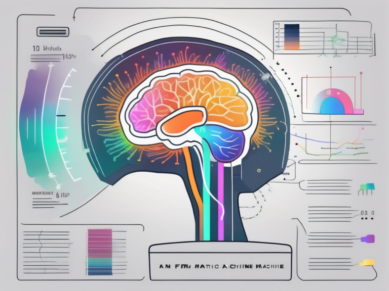
fMRI relies on the relationship between neural activity, oxygen consumption, and blood flow. When a specific brain area becomes active, it requires more oxygen. As a result, blood flow to that area increases to meet the demand. By detecting these changes in blood oxygenation, fMRI can identify brain regions involved in specific tasks or functions.
Furthermore, fMRI has been instrumental in studying various neurological and psychological conditions, such as Alzheimer’s disease, schizophrenia, and depression. By pinpointing the regions of the brain that are affected in these conditions, researchers can develop targeted interventions and treatments to improve patient outcomes.
How Does fMRI Work?
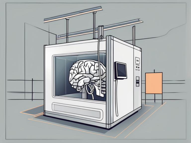
The fMRI process involves the use of a powerful magnetic field and radio waves. When a participant is inside the fMRI scanner, the magnetic field aligns the protons in their body with the field. The radio waves then perturb these aligned protons, and when they return to their original state, they emit signals that are detected by the scanner. These signals are then translated into visual representations of brain activity.
It is important to note that fMRI technology continues to evolve, with ongoing research focused on enhancing the spatial and temporal resolution of brain imaging. These advancements hold promise for uncovering new insights into the complexities of the human brain and could potentially lead to breakthroughs in fields such as cognitive psychology, neurology, and artificial intelligence.
The Role of fMRI in Studying Human Movement
The Brain-Movement Connection
Human movement is a fascinating process that involves a sophisticated interplay between sensory input, motor planning, and execution. The brain is at the core of this intricate mechanism, orchestrating the coordination of various processes to produce fluid and purposeful movements. fMRI, or functional magnetic resonance imaging, serves as a powerful tool for researchers to delve into the neural underpinnings of movement and unravel the complex network of brain regions involved in this fundamental human ability.
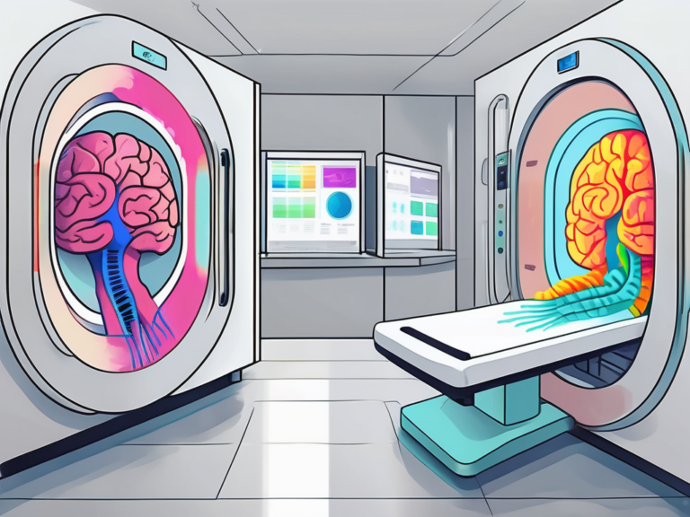
Through the lens of fMRI technology, scientists can pinpoint specific brain regions that are activated during different types of movements, shedding light on the neural circuits responsible for coordinating actions. This deeper understanding of the brain-movement connection not only enhances our knowledge of human physiology but also holds promising implications for fields such as rehabilitation and sports science.
fMRI and Motor Planning
Motor planning is a crucial cognitive process that precedes the execution of a movement, involving the intricate coordination of neural signals to map out the desired action. Within the realm of fMRI studies, researchers have uncovered the intricate dance between brain regions involved in motor planning, highlighting the dynamic interplay between areas such as the prefrontal cortex and parietal cortex. These findings not only deepen our comprehension of how the brain prepares for movement but also pave the way for potential therapeutic interventions targeting motor planning deficits.
Furthermore, the application of fMRI in investigating motor planning offers a window into the complexities of movement disorders and neurodegenerative conditions, providing valuable insights that could inform personalized treatment strategies and rehabilitation protocols.
Decoding Movement Signals with fMRI
Advancements in fMRI technology have opened up new frontiers in the realm of movement research, allowing scientists to decode intricate movement-related information directly from brain activity patterns. By harnessing the power of machine learning algorithms and neural decoding techniques, researchers can now predict specific movements or reconstruct perceived actions based on fMRI data. This groundbreaking capability not only fuels our understanding of the brain’s inner workings but also holds immense potential for innovative applications in the realm of neuroprosthetics.
The ability to decode movement signals from fMRI data has paved the way for cutting-edge developments in brain-computer interfaces, offering hope for individuals with motor impairments to regain autonomy and control over their environment through the power of their thoughts. This transformative technology stands at the forefront of bridging the gap between neuroscience and real-world applications, heralding a new era of possibilities for enhancing human movement capabilities.
The Process of Using fMRI to Study Movement
Preparing for an fMRI Scan
Prior to an fMRI scan, participants are typically informed about the procedure and screened for any contraindications. They are advised to avoid consuming certain substances, such as caffeine or medication that may interfere with the scan. Additionally, participants may be asked to complete specific tasks during the scan, such as performing simple motor movements or imagining movements.
Furthermore, before the fMRI scan, participants are often fitted with specialized equipment to monitor their physiological responses during the procedure. This equipment can include heart rate monitors, respiratory belts, and pulse oximeters. These tools help researchers track changes in vital signs that may influence the fMRI results, ensuring a more accurate interpretation of the data.
The fMRI Scanning Process
During an fMRI scan, participants lie inside the scanner and are asked to remain as still as possible to minimize motion artifacts. The scanner generates a series of images capturing changes in blood flow throughout the brain. These images are then analyzed to identify brain regions associated with movement or motor planning.
In addition to remaining still, participants may also be instructed to follow specific instructions or cues presented to them during the scan. These cues can range from visual prompts on a screen to auditory signals delivered through headphones. By following these instructions, participants help researchers correlate brain activity with specific movements or cognitive tasks, providing valuable insights into the neural mechanisms underlying motor control.
Interpreting fMRI Data
Interpreting fMRI data requires specialized knowledge and expertise. Researchers use sophisticated statistical techniques to analyze the data and identify brain regions that are significantly associated with the task or movement being studied. Care must be taken to distinguish between correlations and causation and to consider various confounding factors that could influence the results.
Moreover, the interpretation of fMRI data often involves collaboration between experts in neuroscience, psychology, and data analysis. By pooling their diverse skill sets, researchers can ensure a comprehensive and nuanced understanding of the complex patterns of brain activity revealed by fMRI scans. This interdisciplinary approach is crucial for drawing accurate conclusions from fMRI studies and advancing our knowledge of how the brain controls movement and behavior.
Challenges and Limitations of Using fMRI
Functional Magnetic Resonance Imaging (fMRI) has revolutionized the field of neuroscience by allowing researchers to non-invasively observe brain activity in real-time. However, like any scientific tool, fMRI comes with its own set of challenges and limitations that researchers must navigate to ensure the validity of their findings.
Potential Drawbacks of fMRI
One of the primary limitations of fMRI is the indirect measurement of neural activity through changes in blood flow. While blood flow is closely linked to neural activity, it is not a direct measure, leading to potential inaccuracies in interpreting brain function. Furthermore, factors such as participant motion, scanner noise, and the confined space of the MRI machine can introduce artifacts that may compromise the quality and reliability of fMRI data.
Another consideration is the issue of reproducibility in fMRI studies. Due to the complex nature of analyzing fMRI data and the multitude of variables that can influence results, replication of findings can sometimes be challenging. This highlights the importance of robust experimental design and transparent reporting to ensure the credibility of fMRI research.
Addressing the Limitations of fMRI
To overcome the challenges associated with fMRI, researchers have developed various strategies and techniques to enhance the accuracy and reliability of their results. Motion correction algorithms are commonly used to compensate for participant movement during scanning, minimizing potential distortions in the data. Additionally, advancements in signal processing and statistical analysis methods continue to refine the interpretation of fMRI signals, improving the specificity of neural activity mapping.
Collaboration between multidisciplinary teams, including neuroscientists, physicists, and data scientists, is essential for advancing fMRI research and addressing its limitations. By combining expertise from diverse fields, researchers can develop innovative approaches to overcome challenges and push the boundaries of our understanding of the human brain.
The Future of Movement Studies with fMRI
Advances in fMRI Technology
Technological advancements continue to propel the field of fMRI forward. High field scanners provide improved spatial and temporal resolution, enabling researchers to visualize fine-grained details of brain activity. For instance, these scanners can now capture the subtle nuances of neural activity during complex movements, such as playing a musical instrument or performing a dance routine. This level of detail allows scientists to gain a deeper understanding of the intricate neural processes involved in these activities.
Additionally, innovative techniques, such as real-time fMRI, are being developed to provide dynamic feedback and facilitate neurofeedback training. Real-time fMRI allows individuals to see their brain activity in real-time, providing them with the opportunity to modulate their neural responses. This technique holds great promise for movement studies, as it can potentially be used to enhance motor learning and rehabilitation. Imagine a future where individuals with movement disorders can use real-time fMRI to train their brains to regain control over their movements, leading to significant improvements in their quality of life.
Potential Future Applications of fMRI in Movement Studies
The future holds exciting possibilities for fMRI in movement studies. Researchers envision using fMRI to better understand and alleviate movement disorders. By studying the neural activity associated with these disorders, scientists can identify potential targets for therapeutic interventions. This knowledge can pave the way for the development of innovative treatments that specifically target the underlying neural mechanisms of movement disorders, offering new hope for patients.
Furthermore, fMRI has the potential to enhance sports performance. By examining the brain activity of elite athletes during different movements, researchers can identify the neural signatures associated with optimal performance. This information can then be used to develop targeted training programs that optimize neural activation patterns, leading to improved athletic performance. Imagine a future where athletes can undergo personalized fMRI assessments to identify their unique neural strengths and weaknesses, allowing them to tailor their training regimens accordingly.
Moreover, fMRI can be used to investigate the neural basis of dance or other complex movements. By studying the brains of professional dancers, researchers can unravel the intricate neural networks that underlie their exceptional motor skills. This knowledge can not only enhance our understanding of the art of dance but also provide valuable insights into motor learning and control in general.
As the field continues to grow, collaborations between scientists, engineers, and clinicians will further expand the potential applications of fMRI in movement research. By combining expertise from various disciplines, researchers can tackle complex challenges and develop innovative solutions that push the boundaries of our understanding of human movement.
In conclusion, fMRI is a valuable tool for understanding how movements are planned and executed in the human brain. By leveraging the relationship between neural activity and blood flow, fMRI provides insights into motor planning, the coordination of movements, and the brain regions involved. However, it is essential to acknowledge the limitations of fMRI and continually strive for advancements in technology and data analysis techniques. As we unlock the full potential of this remarkable imaging modality, we are poised to gain a deeper understanding of human movement and its underlying neural mechanisms.