how are pet and fmri similar
In the field of medical diagnostics, two imaging techniques have revolutionized the way we understand the human body: positron emission tomography (PET) and functional magnetic resonance imaging (fMRI). Although these technologies differ in their underlying principles, they share several similarities that make them invaluable tools for medical professionals. This article aims to delve into the intricacies of both PET and fMRI, exploring their similarities, applications, advantages, limitations, and future developments.
Understanding PET and fMRI Technologies
Defining PET: A Brief Overview
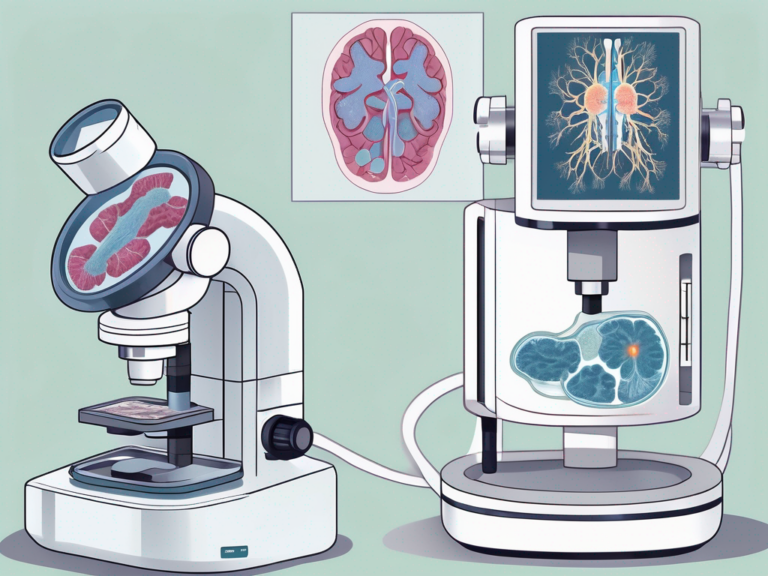
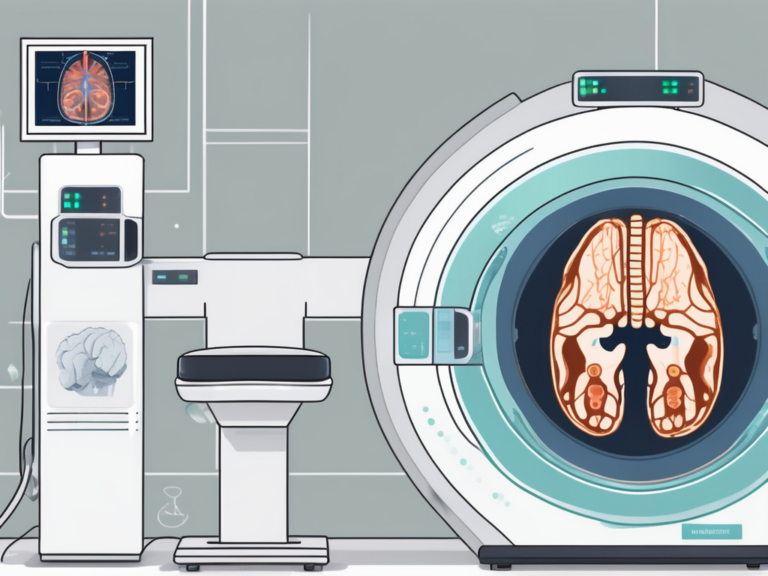
Positron emission tomography, commonly known as PET, is a non-invasive imaging technique that utilizes a special type of radiopharmaceutical tracer to provide detailed physiological information about different organs and tissues within the body. By measuring the emission of positrons, PET scans reveal the metabolic activity and blood flow in specific regions of interest.
PET scans have revolutionized the field of medicine by enabling clinicians to visualize and quantify various biological processes, such as glucose metabolism, neurotransmitter activity, and receptor density. This valuable information helps in the early detection and monitoring of diseases like cancer, Alzheimer’s, and Parkinson’s. Moreover, PET imaging plays a crucial role in neurology, cardiology, and oncology, providing insights into the underlying mechanisms of different conditions.
Defining fMRI: A Brief Overview
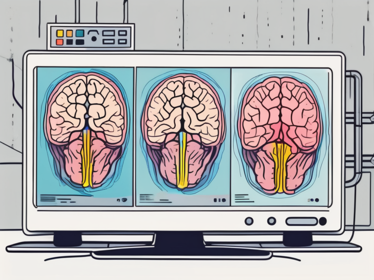
On the other hand, functional magnetic resonance imaging, or fMRI, relies on the powerful magnetic fields of MRI scanners to detect changes in blood oxygenation levels in various brain regions. These changes serve as indirect markers of neuronal responsiveness, allowing researchers to investigate brain function and activity.
fMRI has significantly advanced our understanding of cognitive processes, such as attention, memory, language, and decision-making. By mapping brain activity in real-time, fMRI studies have shed light on how different regions of the brain interact and communicate during various tasks and stimuli. This technology has been instrumental in unraveling the complexities of the human brain and has applications in psychology, neuroscience, and even marketing research.
The Science Behind PET and fMRI
The Physics of PET Scans
In PET scans, the radiopharmaceutical tracer is injected into the patient’s bloodstream. Once inside the body, the tracer releases positively charged particles called positrons, which annihilate upon encountering negatively charged electrons. This annihilation produces gamma rays, which PET detectors capture and use to create three-dimensional images of the tracer distribution. By analyzing these images, healthcare professionals can identify abnormalities or pathological conditions with remarkable precision.
PET scans are particularly useful in oncology, as they can detect cancerous tumors and track the effectiveness of cancer treatments over time. Additionally, PET imaging is crucial in neurology for diagnosing conditions such as Alzheimer’s disease, epilepsy, and traumatic brain injuries. The ability of PET scans to visualize metabolic processes at the molecular level provides invaluable information for both research and clinical applications.
The Physics of fMRI Scans
Contrary to PET scans, fMRI measures the blood oxygen level-dependent (BOLD) signal to infer neuronal activity. When a specific brain region becomes more metabolically active, it requires increased blood flow. The fMRI scanner can detect these changes by monitoring the varying magnetic properties of oxygenated and deoxygenated blood. By mapping the BOLD signal, researchers gain insights into the areas of the brain responsible for certain cognitive processes or responses to external stimuli.
fMRI technology has revolutionized the field of cognitive neuroscience by allowing researchers to observe brain activity in real-time. Through fMRI studies, scientists have made significant discoveries about how the brain processes information, forms memories, and responds to emotional stimuli. This non-invasive imaging technique has opened up new avenues for understanding complex neurological disorders such as schizophrenia, depression, and autism spectrum disorders.
Similarities in Imaging Techniques
The Use of Radioactive Tracers in Both Techniques
Both PET and fMRI techniques rely on the use of specialized tracers. While PET employs radiopharmaceuticals, fMRI utilizes contrast agents that alter the magnetic properties of blood. These tracers enable healthcare providers and researchers to visualize and analyze specific physiological processes, contributing to the diagnosis and understanding of various conditions.
Furthermore, the use of radioactive tracers in imaging techniques has revolutionized the field of medicine by allowing for non-invasive visualization of internal structures and functions. Radiopharmaceuticals used in PET scans are carefully selected to target specific organs or tissues, providing valuable insights into metabolic processes and disease progression. On the other hand, contrast agents in fMRI help highlight areas of increased neural activity in the brain, aiding in the study of cognitive functions and neurological disorders.
The Role of Magnetic Fields in Imaging
Magnetic fields play an essential role in both PET and fMRI scans. PET detectors rely on magnetic fields to accurately measure and capture the gamma rays emitted during positron-electron annihilation. Similarly, fMRI scanners utilize powerful and precisely controlled magnetic fields to create detailed images of brain activity based on the BOLD signal.
Moreover, the magnetic fields used in imaging techniques are carefully calibrated to ensure optimal signal-to-noise ratios and spatial resolution. In PET imaging, the magnetic fields help reduce background noise and improve the accuracy of gamma ray detection, leading to clearer and more precise images. In fMRI, the magnetic fields are crucial for aligning the magnetic moments of hydrogen atoms in the blood, allowing for the detection of changes in blood oxygenation levels that correspond to neural activity.
Applications of PET and fMRI in Medical Diagnostics
PET in Oncology and Neurology
PET scans have proven particularly valuable in oncology and neurology. In oncology, PET is frequently used to identify and stage various types of cancer, monitor treatment response, and detect early signs of disease recurrence. In neurology, PET plays a significant role in diagnosing and understanding neurodegenerative disorders such as Alzheimer’s disease, Parkinson’s disease, and epilepsy.
Furthermore, PET imaging is not limited to oncology and neurology. It is also utilized in cardiology to assess myocardial viability, in psychiatry to study various mental health conditions, and in infectious disease to detect sites of infection and monitor treatment progress. The versatility of PET technology highlights its widespread application in diverse medical specialties.
fMRI in Brain Mapping and Stroke Detection
fMRI has revolutionized the field of brain mapping, enabling researchers to identify regions of the brain involved in specific cognitive functions or sensory processing. Moreover, fMRI has shown promise in the timely detection and diagnosis of strokes by quickly identifying compromised brain areas, aiding in the delivery of lifesaving interventions.
Additionally, fMRI is instrumental in studying psychiatric disorders such as schizophrenia and depression, providing insights into the neural mechanisms underlying these conditions. It is also used in the assessment of traumatic brain injuries, helping clinicians understand the impact of such injuries on brain function and guiding rehabilitation strategies. The non-invasive nature of fMRI makes it a valuable tool in both research and clinical settings, offering detailed insights into brain structure and function.
Advantages and Limitations of PET and fMRI
Strengths of PET Scans
PET scans offer unparalleled sensitivity in detecting and characterizing metabolic abnormalities, making them ideal for cancer staging and monitoring treatment effectiveness. Additionally, PET tracers can target specific biochemical processes, providing insights into the underlying molecular mechanisms of diseases.
For example, in oncology, PET scans can help determine the extent of tumor spread by visualizing areas of increased glucose metabolism. This information is crucial for accurate staging, treatment planning, and assessing response to therapy. Moreover, PET tracers such as fluorodeoxyglucose (FDG) can reveal metabolic changes in the brain associated with neurodegenerative disorders like Alzheimer’s disease, aiding in early diagnosis and monitoring disease progression.
Strengths of fMRI Scans
fMRI excels in spatial resolution, allowing for precise localization of brain activity. The non-invasive nature of fMRI also makes it a safer alternative to invasive techniques such as electrode implantation. Moreover, fMRI studies can be designed to investigate dynamic changes in brain activity over time, providing valuable information about cognitive processes.
For instance, fMRI has been instrumental in unraveling the neural correlates of memory formation. By examining the activation patterns in different brain regions during memory tasks, researchers have gained insights into the intricate network of brain regions involved in encoding, storage, and retrieval of memories. This knowledge has implications not only for understanding normal cognitive processes but also for developing interventions to treat memory disorders.
Limitations of PET Scans
One inherent limitation of PET is its reliance on radiopharmaceutical tracers with a short half-life, which limits the availability and practicality of certain tracers for clinical use. PET scans also involve exposure to ionizing radiation, albeit at minimal levels. Finally, the cost of PET scans and the need for specialized equipment can be potential drawbacks.
Furthermore, the interpretation of PET scan results requires expertise in image analysis and knowledge of the specific tracer used. This can pose challenges in clinical settings where access to specialized personnel may be limited. Despite these limitations, PET remains a valuable tool in various fields of medicine, providing unique insights into the metabolic activity of tissues and organs.
Limitations of fMRI Scans
fMRI is sensitive to motion artifacts, meaning even subtle head movements during the scan can introduce errors in the results. Consequently, fMRI studies often require participants to remain still for extended periods, which may limit the generalizability of findings. Additionally, fMRI primarily measures changes in blood flow, introducing a delay in the signal that can complicate the interpretation of findings.
Moreover, fMRI has inherent limitations in its ability to directly measure neural activity. The blood oxygen level-dependent (BOLD) signal, which fMRI relies on, is an indirect measure of neuronal activity and is subject to various confounding factors. These factors include fluctuations in blood pressure, carbon dioxide levels, and cerebral autoregulation. Researchers must carefully consider these limitations when drawing conclusions from fMRI studies.
The Future of PET and fMRI Technologies
Innovations in PET Imaging
Ongoing research aims to advance PET technologies, particularly in terms of radiotracer development and imaging resolution. New radiotracers with longer half-lives and greater affinity for specific molecular targets will expand the diagnostic capabilities of PET, facilitating the early detection and personalized treatment of various diseases. Furthermore, technological advancements may lead to faster and more precise PET scanners, reducing imaging times and improving patient comfort.
One exciting area of research in PET imaging is the development of novel radiotracers that can target specific cellular processes involved in disease progression. For example, scientists are currently exploring radiotracers that can detect and quantify the activity of specific enzymes that play a crucial role in cancer growth. By visualizing the activity of these enzymes, PET imaging can provide valuable information about tumor aggressiveness and response to treatment, enabling physicians to tailor therapies to individual patients.
Innovations in fMRI Imaging
fMRI studies continue to evolve, with researchers harnessing machine learning algorithms to decipher complex patterns of brain activity. This integration of artificial intelligence and fMRI opens up exciting new possibilities, facilitating the identification of subtle brain alterations and aiding early detection of neurological disorders. Additionally, advancements in fMRI hardware and real-time processing will enhance imaging capabilities, allowing for unprecedented insights into brain function and connectivity.
Another fascinating area of research in fMRI imaging is the exploration of brain networks and their role in various cognitive processes. By analyzing the coordinated activity of different brain regions, researchers can gain a deeper understanding of how the brain functions as a complex network. This knowledge has the potential to revolutionize our understanding of neurological disorders such as Alzheimer’s disease, schizophrenia, and depression, by uncovering the underlying network disruptions that contribute to these conditions.
As the fields of PET and fMRI continue to intersect and complement each other, their similarities become increasingly evident. Together, these imaging techniques provide valuable information about the body’s inner workings, aiding in the diagnosis, treatment, and understanding of numerous medical conditions. While each technique has its strengths and limitations, ongoing advancements promise to overcome current challenges, ushering in an era of even greater precision and accessibility in medical diagnostics.