what does fmri measure
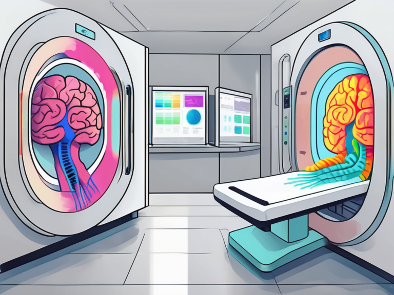
Functional Magnetic Resonance Imaging, commonly known as fMRI, is a powerful technique used in neuroscience to measure brain activity. By detecting changes in blood flow, fMRI provides valuable insights into brain functions and the underlying neural processes. In this article, we will explore the basics of fMRI, its key components, the process of measuring with fMRI, what fMRI measures specifically, applications of fMRI in neuroscience, its limitations and challenges, and the future of this exciting field.
Understanding the Basics of fMRI
Functional Magnetic Resonance Imaging relies on the principles of magnetic resonance, which allows us to examine the living brain in a non-invasive manner. One of the key advantages of fMRI is its high spatial resolution, enabling researchers to pinpoint brain activity at a detailed level. Before diving into the specifics, it is crucial to understand the science behind fMRI and its key components.
The Science Behind fMRI
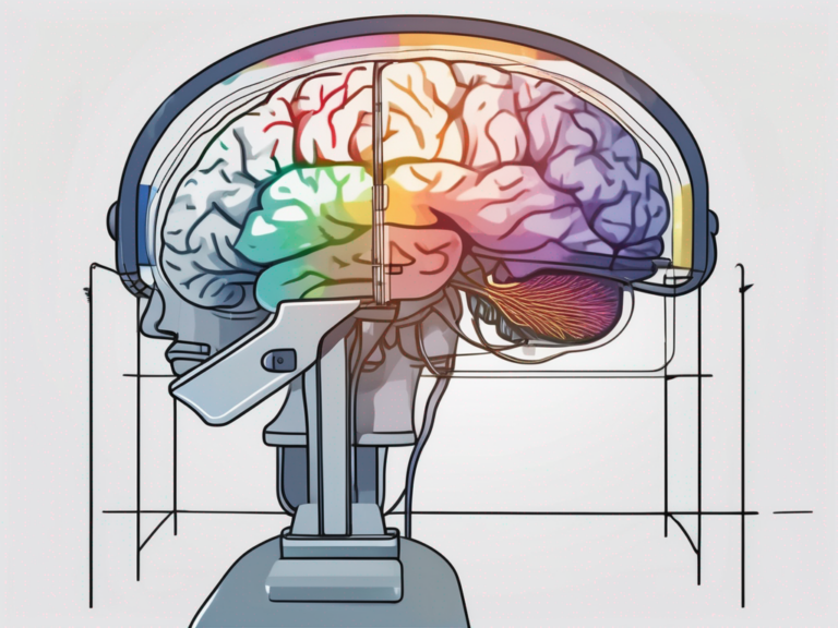
At the heart of fMRI lies a phenomenon known as the blood-oxygen-level-dependent (BOLD) contrast. When a particular brain region becomes active, it requires more oxygenated blood. This increased demand for oxygen triggers a cascading effect, resulting in changes in blood flow and oxygenation in the active area. By capturing these changes, fMRI provides a window into brain activity.
Moreover, fMRI not only reveals which areas of the brain are active during specific tasks but also allows researchers to observe how different regions communicate with each other. This connectivity information is crucial for understanding complex brain functions such as memory, decision-making, and emotions.
Key Components of an fMRI Machine
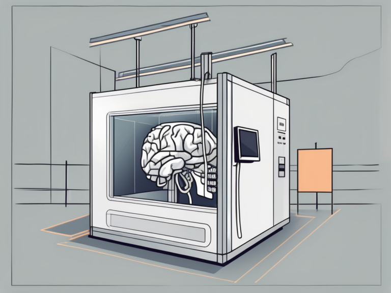
An fMRI machine consists of several essential components, including a powerful magnet, radiofrequency coils, and a computer system. The magnet creates a strong magnetic field that aligns the protons in the human body. The radiofrequency coils emit radio waves, which stimulate the protons to emit signals. These signals are captured by the machine and processed to generate the fMRI images we analyze.
Additionally, the computer system in an fMRI machine plays a vital role in processing the raw data collected during a scan. Sophisticated algorithms are used to convert the signals into detailed images of brain activity. These images are then carefully analyzed by researchers to draw meaningful conclusions about the cognitive processes underlying the tasks performed by the study participants.
The Process of Measuring with fMRI
Now that we understand the basics, let’s delve into the process of measuring brain activity with fMRI. From preparing for an fMRI scan to the role of magnetic fields in fMRI, this section will guide you through the necessary steps involved in obtaining valuable fMRI data.
Functional Magnetic Resonance Imaging (fMRI) is a powerful tool used in neuroscience to measure brain activity by detecting changes in blood flow. This non-invasive technique provides researchers with valuable insights into how different regions of the brain are involved in various cognitive tasks and functions.
Preparing for an fMRI Scan
Prior to an fMRI scan, patients are typically instructed to remove any metal objects and change into appropriate clothing without metal attachments. This is necessary since the powerful magnet in the fMRI machine can interact with metallic objects and cause harm. Additionally, patients are informed about the procedure and given the opportunity to address any concerns they may have.
Furthermore, patients are often asked to remain still during the scan to ensure clear imaging results. Any movement can distort the images and affect the accuracy of the data collected. To help patients stay comfortable and reduce anxiety, some fMRI facilities offer headphones to listen to music or communicate with the technician during the scan.
The Role of Magnetic Fields in fMRI
Magnetic fields play a fundamental role in the functioning of fMRI. The strong magnetic field generated by the machine aligns the protons in the brain, allowing them to emit signals. By manipulating these signals, the fMRI machine creates detailed images that highlight changes in blood flow and oxygenation, reflecting brain activity.
Moreover, the magnetic field strength of an fMRI machine is measured in units called Tesla (T). Higher Tesla machines offer better image resolution and clarity, enabling researchers to capture more detailed information about brain activity. The magnetic field is carefully controlled and calibrated to ensure precise and accurate measurements during the scanning process.
What fMRI Measures: Brain Activity
Now that we have covered the basics of fMRI and the process of measuring with it, let’s explore what exactly fMRI is capable of measuring in terms of brain activity. By detecting changes in blood flow and oxygenation, fMRI offers valuable insights into the functioning of the human brain.
Functional Magnetic Resonance Imaging (fMRI) is a powerful tool used in neuroscience to study brain function. It works by detecting changes in blood flow and oxygenation levels in the brain, providing researchers with a way to indirectly measure neural activity. This non-invasive technique has revolutionized our understanding of the brain and its intricate workings.
How fMRI Detects Changes in Blood Flow
fMRI relies on the BOLD contrast, which detects changes in blood flow that accompany brain activity. When neurons become active, they consume more energy in the form of oxygen. In response, nearby blood vessels dilate to deliver oxygen-rich blood to the active region. This increase in blood flow and oxygenation is precisely what fMRI measures, providing an indirect indication of brain activity.
Moreover, fMRI can differentiate between oxygenated and deoxygenated blood, allowing researchers to map out specific brain regions that are involved in various cognitive processes. This level of detail helps in understanding the neural mechanisms underlying complex behaviors and mental functions.
Interpreting fMRI Data
Interpreting fMRI data involves analyzing the patterns of brain activity captured by the machine. Researchers employ sophisticated statistical techniques to identify regions that show increased activity in response to certain tasks or stimuli. These findings have implications for various fields, from cognitive studies to clinical diagnosis and treatment.
Furthermore, fMRI data can be used in conjunction with other neuroimaging techniques, such as EEG and PET scans, to provide a more comprehensive understanding of brain function. This multi-modal approach allows researchers to investigate brain activity at different spatial and temporal scales, leading to more nuanced insights into the complexities of the human brain.
Applications of fMRI in Neuroscience
fMRI has revolutionized our understanding of the human brain and its functions. This versatile technique finds applications in numerous areas of research, contributing to our knowledge of cognitive processes, disease mechanisms, and potential therapeutic interventions.
One fascinating aspect of fMRI is its ability to provide insights into the brain’s intricate network of connections. By measuring changes in blood flow, researchers can identify regions of the brain that are activated during specific tasks or stimuli. This information is crucial for understanding how different brain regions communicate and work together to produce complex behaviors and thoughts.
Cognitive Studies Using fMRI
fMRI allows researchers to investigate the neural correlates of cognitive processes, such as attention, memory, language, and decision-making. By examining brain activity during specific tasks or stimuli, scientists can unravel the underlying mechanisms and shed light on the complexities of the human mind.
Furthermore, fMRI studies have revealed the plasticity of the brain, showing how neural circuits can reorganize and adapt in response to learning, experience, and injury. This phenomenon highlights the brain’s remarkable ability to change and adapt throughout life, offering hope for novel therapeutic approaches for various neurological conditions.
fMRI in Clinical Diagnosis and Treatment
fMRI has found applications in clinical settings, aiding in the diagnosis and treatment of various neurological and psychiatric conditions. By mapping brain activity, fMRI can help identify abnormalities and guide treatment strategies, leading to personalized and more effective interventions.
Moreover, fMRI is increasingly being used in pre-surgical planning for patients with brain tumors or epilepsy. By mapping out critical brain regions involved in essential functions such as movement, language, and sensation, surgeons can better plan their procedures to minimize potential damage and improve patient outcomes. This integration of fMRI into clinical practice represents a significant advancement in personalized medicine and neurosurgical care.
Limitations and Challenges of fMRI
Like any scientific technique, fMRI has its limitations and faces certain challenges. Understanding these drawbacks is crucial for accurate interpretation and cautious application.
One significant limitation of fMRI is its temporal resolution, which refers to the ability to precisely capture the timing of neural activity. While fMRI provides detailed spatial information about brain regions involved in specific tasks or stimuli, it has a relatively poor temporal resolution compared to techniques like electroencephalography (EEG) or magnetoencephalography (MEG). This limitation makes it challenging to track the rapid dynamics of neural processes, especially those occurring within milliseconds.
Potential Risks and Side Effects
fMRI is generally considered safe. However, patients with certain conditions and medical devices, such as pacemakers or cochlear implants, may be at risk due to the strong magnetic fields involved. Additionally, some individuals may experience anxiety or claustrophobia during fMRI scans, highlighting the importance of appropriate pre-scan preparations.
Another challenge in fMRI research is the issue of signal-to-noise ratio (SNR), which can affect the quality and reliability of the data obtained. The fMRI signal is relatively weak compared to noise sources such as physiological fluctuations, motion artifacts, and scanner-related distortions. Researchers employ various techniques, such as signal processing algorithms and noise reduction methods, to enhance the SNR and improve the accuracy of fMRI results.
Controversies and Criticisms of fMRI
Despite its many advancements and applications, fMRI has faced criticism and controversies within the scientific community. Some argue that the statistical methods employed in fMRI data analysis can lead to false positives or overinterpretation. Others question the reproducibility and generalizability of fMRI findings. Ongoing research aims to address these concerns and improve the reliability of fMRI studies.
Furthermore, the issue of neurovascular coupling, which refers to the relationship between neural activity and cerebral blood flow, is a topic of ongoing debate in fMRI research. The assumption that changes in blood flow accurately reflect neural activity has been challenged, leading to discussions about the complex interplay between neuronal and vascular responses measured by fMRI. Understanding the intricacies of neurovascular coupling is essential for the accurate interpretation of fMRI data and the advancement of neuroimaging techniques.
The Future of fMRI
The field of functional Magnetic Resonance Imaging (fMRI) continues to be at the forefront of neuroscience research, driving significant advancements in our understanding of the human brain. As technology and methodologies progress, the future of fMRI holds great promise for unraveling the mysteries of the mind.
One exciting area of development in fMRI technology is the exploration of novel imaging sequences and pulse sequences that can provide even higher spatial and temporal resolution. These advancements not only offer more detailed insights into brain activity but also open up new possibilities for studying dynamic processes within the brain with unprecedented clarity.
Innovations in fMRI Technology
Researchers are also delving into the realm of multi-modal imaging, combining fMRI with techniques such as electroencephalography (EEG) and magnetoencephalography (MEG) to complement each other’s strengths and provide a more comprehensive view of brain function. By integrating these different modalities, scientists can overcome the limitations of each individual technique and gain a more holistic understanding of brain activity.
Furthermore, the integration of fMRI with virtual reality environments is revolutionizing cognitive neuroscience research by allowing for more ecologically valid experiments. This merging of technology creates immersive scenarios that closely mimic real-world experiences, enabling researchers to study brain responses in a more naturalistic setting.
Emerging Trends in fMRI Research
As fMRI research progresses, there is a growing emphasis on studying brain connectivity and network dynamics to uncover the intricate relationships between different regions of the brain. By analyzing large-scale brain networks, researchers can gain insights into how information is processed and integrated across various brain regions, shedding light on the underlying mechanisms of cognition and behavior.
Moreover, the field of fMRI is increasingly embracing open science practices, promoting data sharing and collaboration to accelerate scientific discoveries. By fostering transparency and reproducibility in research, the fMRI community aims to build a solid foundation of knowledge that can drive future breakthroughs in understanding the complexities of the human brain.
Conclusion
Functional Magnetic Resonance Imaging is a powerful tool that measures brain activity by detecting changes in blood flow. Understanding the basics of fMRI, its key components, and the process of measuring with it is crucial for conducting meaningful research and interpreting data accurately. The applications, limitations, and future of fMRI hold great promise for advancing our understanding of the human brain and contributing to various fields, from cognitive neuroscience to clinical practice.