why do fmri and eeg still coexist as useful brain scanning techniques?
In the field of neuroscience, there are several techniques available for scanning the human brain. Two widely used and valuable methods are functional magnetic resonance imaging (fMRI) and electroencephalography (EEG). Despite their differences, fMRI and EEG continue to coexist as useful brain scanning techniques due to their unique advantages and complementary nature.
Understanding the Basics of fMRI and EEG
What is fMRI?
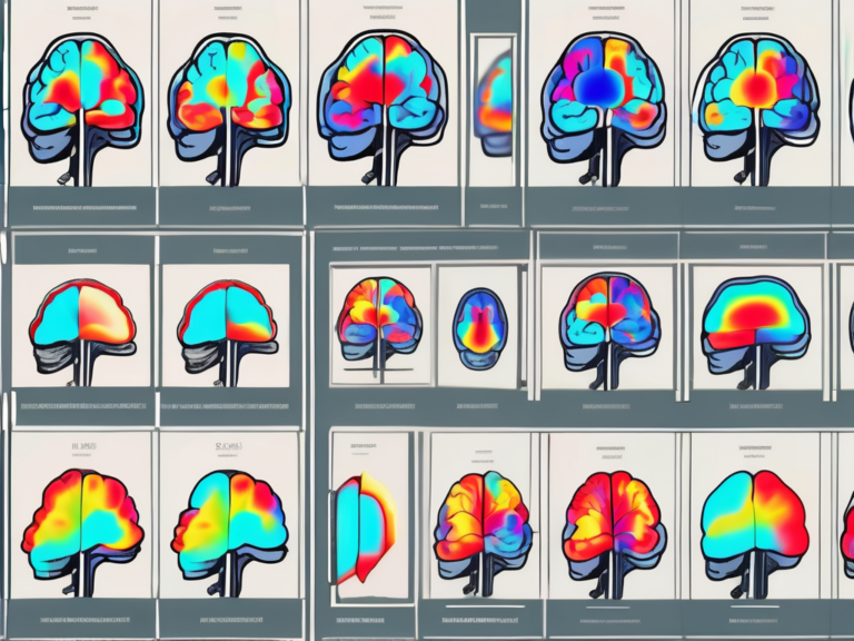
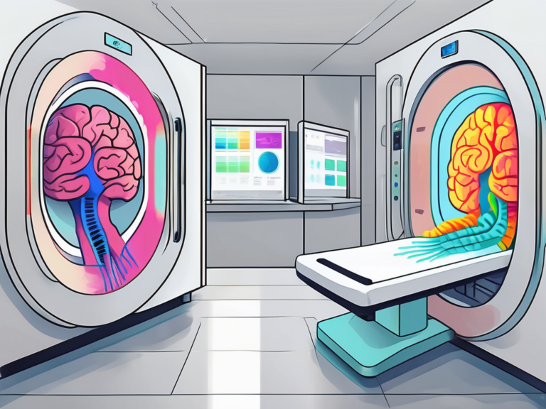
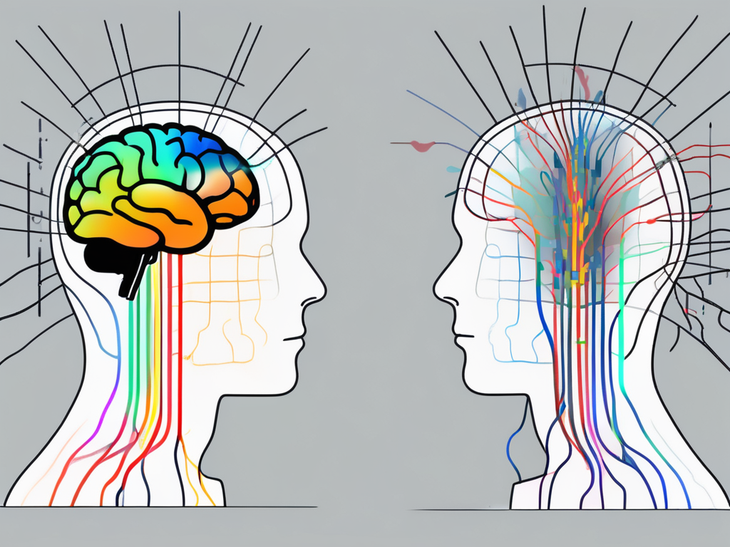
Functional magnetic resonance imaging (fMRI) is a non-invasive imaging technique that measures brain activity by detecting changes in blood flow. This method relies on the fact that when a certain brain region is active, it requires increased oxygen and glucose, resulting in a change in blood flow to that area. By measuring these changes, fMRI can pinpoint the brain regions associated with specific tasks or cognitive processes.
One fascinating aspect of fMRI is its ability to provide both spatial and temporal information about brain activity. Not only can it show which areas of the brain are involved in a particular task, but it can also reveal the sequence in which these areas are activated. This temporal dimension adds a valuable layer of understanding to cognitive processes and helps researchers unravel the complexities of brain function.
What is EEG?
Electroencephalography (EEG) is another non-invasive technique used to measure brain activity. It records the electrical signals produced by the brain using a set of electrodes placed on the scalp. These electrodes detect the collective activity of millions of neurons in the brain, providing an overview of the brain’s electrical rhythms and patterns.
EEG is particularly useful in studying rapid brain processes, such as those involved in perception and motor activities. Its high temporal resolution allows researchers to observe brain activity in real-time, capturing the millisecond-by-millisecond changes that occur during various cognitive tasks. This real-time monitoring is crucial for understanding the dynamics of brain function and how different regions of the brain communicate with each other during different activities.
The Unique Advantages of fMRI
High Spatial Resolution
One of the notable advantages of fMRI is its high spatial resolution. It can precisely locate brain activity within millimeter-sized voxels, allowing researchers to identify specific brain regions involved in particular tasks or functions. This level of detail is crucial for understanding the localization of cognitive processes and mapping brain networks.
Furthermore, the high spatial resolution of fMRI enables researchers to investigate subtle differences in brain activity that may not be detectable with other imaging techniques. This precision is particularly valuable in studying complex brain disorders where pinpointing specific brain regions involved is essential for developing targeted treatments.
Non-Invasive Technique
fMRI is a non-invasive procedure, which means it does not require any direct contact with the brain. Unlike invasive techniques such as implanting electrodes, fMRI offers a safe and comfortable experience for participants. This non-invasiveness makes it an appealing option for studying various populations, including children and individuals with certain medical conditions.
Moreover, the non-invasive nature of fMRI allows for longitudinal studies where participants can be scanned multiple times without the risks associated with invasive procedures. This capability is invaluable for tracking changes in brain activity over time, providing insights into the progression of neurological disorders or the effects of interventions.
The Distinct Benefits of EEG
Superior Temporal Resolution
EEG excels in providing superior temporal resolution. It can capture the brain’s electrical activity in real-time, with millisecond precision. This high temporal resolution enables researchers to analyze rapid changes in brain activity and explore the dynamic nature of cognitive processes, such as attention, perception, and memory.
Furthermore, the millisecond precision of EEG is crucial in studying event-related brain responses, where researchers can pinpoint the exact timing of neural processes in response to specific stimuli. This level of detail is essential in understanding the intricate neural mechanisms underlying various cognitive functions and behaviors.
Accessibility and Affordability
EEG equipment, compared to fMRI machines, is more accessible and affordable. This accessibility allows researchers and clinicians to conduct studies and diagnose certain conditions with greater convenience. Moreover, portable EEG devices have emerged, facilitating the monitoring of brain activity in various environments, including home-based settings.
Additionally, the affordability of EEG equipment has paved the way for its use in diverse fields beyond neuroscience, such as brain-computer interface research, sleep studies, and even entertainment (e.g., brain-controlled games). This versatility highlights the widespread impact of EEG technology in both scientific and commercial applications.
The Complementary Nature of fMRI and EEG
fMRI and EEG: A Holistic View of Brain Function
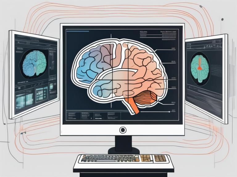
fMRI and EEG provide complementary information about brain function. While fMRI reveals the precise location of brain activity, EEG captures the brain’s electrical activity over time. By combining these two techniques, researchers can obtain a more comprehensive understanding of how the brain functions as a whole – both in terms of brain regions and the dynamics of neural activity.
Furthermore, the integration of fMRI and EEG data has opened up avenues for exploring brain function in real time. This real-time analysis allows researchers to observe how different brain regions interact and communicate with each other during various cognitive tasks or emotional responses. Understanding these dynamic interactions is crucial for unraveling the complexities of the human brain.
The Synergy between fMRI and EEG
The synergy between fMRI and EEG has led to the development of novel analysis methods, such as simultaneous recordings of both modalities. These joint recordings allow researchers to investigate the relationship between brain function and underlying neural processes more deeply. The integration of fMRI and EEG data has the potential to unlock new insights into brain disorders and neuropsychological phenomena.
Moreover, the combination of fMRI and EEG data has paved the way for advancements in neurofeedback techniques. Neurofeedback, a form of biofeedback that uses real-time displays of brain activity to teach self-regulation, has shown promising results in treating conditions such as ADHD and anxiety disorders. By utilizing the complementary strengths of fMRI and EEG, researchers can tailor neurofeedback interventions to target specific brain networks and optimize therapeutic outcomes.
Current Challenges in Brain Imaging Techniques
Limitations of fMRI
Despite its advantages, fMRI has limitations. It is sensitive to motion artifacts, and even slight movements can affect the quality of the data. This can be particularly challenging when studying populations such as children or individuals with movement disorders. Researchers have developed various techniques to address motion artifacts, such as using specialized headgear or advanced image processing algorithms. Additionally, fMRI measures blood flow indirectly, and the relationship between neural activity and blood flow can be complex, requiring careful interpretation. Understanding the hemodynamic response function and its variations across individuals is crucial for accurate data analysis.
Limitations of EEG
EEG is sensitive to noise from various sources, such as muscle activity and eye movements. These artifacts can obscure the desired electrical signals, making data analysis challenging. To mitigate these issues, researchers often employ signal processing methods like independent component analysis (ICA) to separate noise components from brain signals. Furthermore, the spatial resolution of EEG is relatively lower than fMRI, which makes it difficult to precisely localize brain activity. This limitation is due to the nature of EEG signals, which are influenced by the conductivity of different tissues in the head. Advances in EEG technology, such as high-density electrode arrays and sophisticated source localization algorithms, aim to improve spatial resolution and provide more accurate localization of neural activity.
The Future of Brain Scanning Techniques
Technological Advancements in fMRI and EEG
Ongoing technological advancements continue to enhance the capabilities of both fMRI and EEG. Improvements in fMRI techniques, such as faster image acquisition and higher field strengths, increase spatial resolution and provide more detailed brain maps. Similarly, advancements in EEG hardware and signal processing algorithms enhance signal quality and reduce noise, allowing for better interpretation of brain activity.
Moreover, the development of artificial intelligence (AI) algorithms has revolutionized the analysis of fMRI and EEG data. AI can detect subtle patterns in brain scans that may not be apparent to the human eye, leading to more accurate interpretations of brain activity. This intersection of neuroscience and AI opens up new possibilities for understanding the complexities of the human brain.
The Potential of Combined fMRI and EEG Techniques
The integration of fMRI and EEG holds great promise for the future of brain scanning. Combining the strengths of both techniques can provide a comprehensive understanding of brain function, integrating spatial and temporal aspects. This combined approach may lead to breakthroughs in our understanding of neurological disorders, cognitive processes, and brain-computer interface technologies.
Furthermore, the emerging field of multimodal imaging, which combines fMRI, EEG, and other imaging techniques such as magnetoencephalography (MEG) and positron emission tomography (PET), offers a more holistic view of brain function. By integrating data from multiple sources, researchers can overcome the limitations of individual techniques and gain a more nuanced understanding of brain activity.
In conclusion, the coexistence of fMRI and EEG as useful brain scanning techniques can be attributed to their unique advantages, complementary nature, and potential for future advancements. By harnessing the strengths of both techniques, researchers gain a more comprehensive view of brain function, paving the way for a deeper understanding of the complex workings of the human mind.