how does fmri work
Functional Magnetic Resonance Imaging, commonly known as fMRI, is a powerful tool in the field of neuroscience that allows researchers to better understand the human brain. By capturing detailed images of the brain’s activity, fMRI enables scientists to explore various cognitive processes, study neurological disorders, and develop new treatments. In this article, we will delve into the inner workings of fMRI, explore its applications in medicine and research, discuss its advantages and limitations, and take a glimpse into the future of this remarkable technology.
Understanding the Basics of fMRI
Before we dive into the intricacies of fMRI, let’s establish a solid understanding of its fundamental components. At its core, fMRI operates on the principles of magnetic resonance imaging (MRI) and blood oxygen level-dependent (BOLD) contrast. By measuring changes in blood flow and oxygenation, fMRI can detect brain activity in real-time.
Functional magnetic resonance imaging (fMRI) has revolutionized the field of neuroscience by providing researchers with a powerful tool to investigate the inner workings of the human brain. This non-invasive imaging technique allows scientists to observe brain activity by tracking changes in blood flow and oxygen levels. By mapping out which areas of the brain are activated during specific tasks or stimuli, fMRI has significantly advanced our understanding of cognitive processes, emotions, and neurological disorders.
The Science Behind fMRI
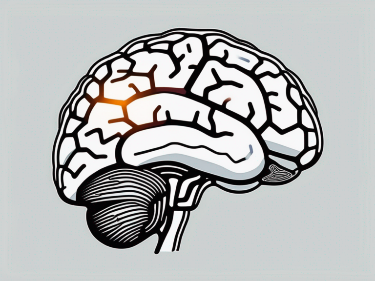
To comprehend how fMRI works, we need to familiarize ourselves with the concept of neurovascular coupling. When a specific area of the brain becomes active, it requires a greater supply of oxygen-rich blood. In response to this increased demand, localized blood vessels dilate, allowing more blood to reach the active region. This intricate relationship between neural activity and blood flow forms the basis of fMRI.
Furthermore, the blood oxygen level-dependent (BOLD) signal detected by fMRI is a complex interplay of physiological processes. Changes in deoxyhemoglobin concentration influence the magnetic properties of blood, leading to alterations in the MRI signal. By analyzing these signal changes, researchers can infer neural activity and create detailed maps of brain function.
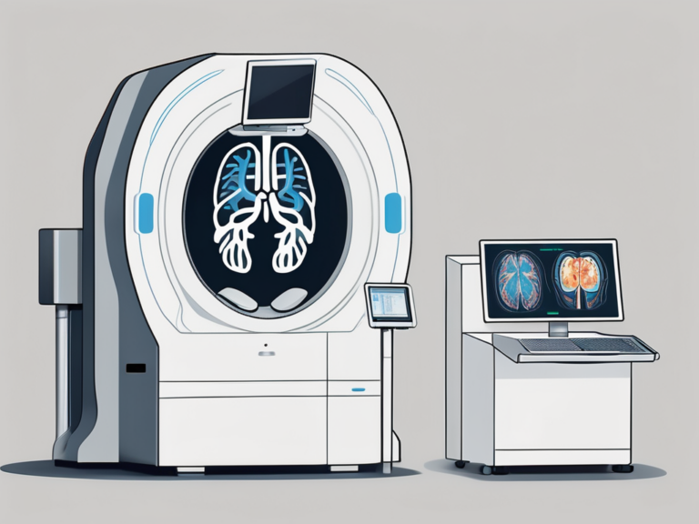
Key Components of an fMRI Machine
An fMRI scanner consists of several vital components that work harmoniously to acquire and process brain images. The primary components include the magnet, radiofrequency coils, gradient coils, and a computer system. The magnet generates a strong magnetic field, while the coils transmit and receive radio waves to capture the brain’s signals. Meanwhile, the gradient coils create a spatial encoding that enables precise localization of brain activity.
Additionally, the fMRI machine requires sophisticated software to reconstruct and analyze the vast amount of data collected during a scan. Advanced algorithms are used to convert raw signals into detailed images of brain activity, allowing researchers to identify specific brain regions involved in different cognitive tasks. The integration of hardware and software in fMRI systems plays a crucial role in unlocking the mysteries of the human brain.
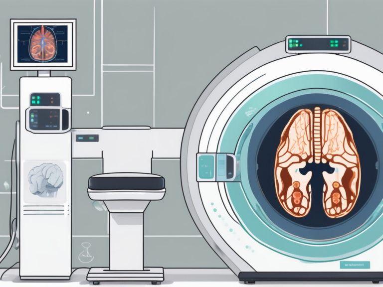
The Process of fMRI Scanning
Now that we have a solid foundation, let’s explore the step-by-step process of undergoing an fMRI scan.
Functional Magnetic Resonance Imaging (fMRI) is a powerful tool used in neuroscience to visualize brain activity in real-time. By measuring changes in blood flow, fMRI can pinpoint which areas of the brain are activated during different tasks or stimuli, providing valuable insights into brain function.
Preparing for an fMRI Scan
Prior to the scan, it’s important to remove all metallic items, such as jewelry or watches, as they can interfere with the magnetic field. The patient is then positioned comfortably on the scanner bed, and a head coil is placed to capture brain signals. It’s crucial to remain as still as possible throughout the scan to ensure optimal image quality.
Additionally, before the scan begins, the patient may undergo a brief practice session to familiarize themselves with the tasks they will be performing inside the scanner. This helps reduce anxiety and ensures accurate data collection during the actual scan.
What Happens During an fMRI Scan
During the scan, a series of images are acquired as the patient performs specific cognitive tasks or rests quietly. These tasks could range from simple actions like tapping fingers to more complex activities like solving puzzles or recalling memories. The scanner’s software analyzes the data and transforms it into detailed images that display the activated brain regions.
As the scan progresses, the patient may hear loud tapping or knocking noises coming from the MRI machine. These sounds are a normal part of the scanning process and are caused by the switching of magnetic fields within the machine. To help mitigate any discomfort from the noise, patients are often provided with earplugs or headphones to wear during the scan.
Interpreting fMRI Results
Now that we have a grasp of how fMRI scans are conducted, let’s explore how researchers interpret the resulting images.
Functional Magnetic Resonance Imaging (fMRI) has revolutionized the field of neuroscience by allowing researchers to observe brain activity in real-time. By measuring changes in blood flow and oxygen levels, fMRI provides valuable insights into which areas of the brain are involved in specific tasks or cognitive processes.
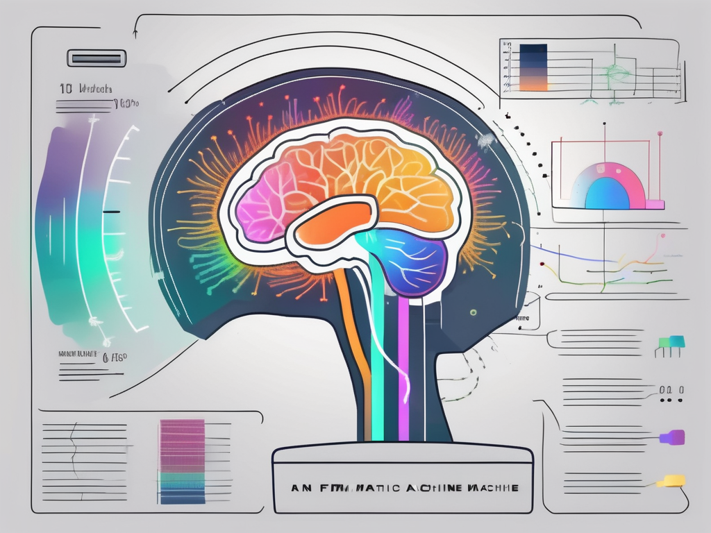
The Role of Color in fMRI Images
One fascinating feature of fMRI images is the use of color to represent brain activity. Typically, researchers use a color scale to indicate variations in blood flow and neural activation. Warmer colors (e.g., red and yellow) signify higher levels of activity, while cooler colors (e.g., blue and green) indicate lower activity.
Color coding in fMRI images not only enhances visual interpretation but also aids in identifying regions of interest within the brain. Researchers can overlay these color-coded images onto anatomical brain scans to pinpoint specific areas that are active during different tasks or stimuli.
Understanding fMRI Data
Interpreting fMRI data requires advanced statistical analysis techniques. Researchers employ statistical models to determine whether the observed brain activity is significantly different from baseline levels. By comparing different conditions or groups, they can unravel the underlying patterns and relationships within the data, providing vital insights into brain function.
Furthermore, fMRI data can be analyzed using various methods such as region of interest (ROI) analysis, voxel-based morphometry (VBM), and functional connectivity analysis. Each of these approaches offers unique perspectives on brain function and connectivity, allowing researchers to delve deeper into the complexities of neural networks and cognitive processes.
Applications of fMRI in Medicine and Research
Beyond its technical aspects, fMRI has revolutionized our understanding of the human brain. Let’s explore some of its remarkable applications in medicine and research.
Functional Magnetic Resonance Imaging (fMRI) is a powerful tool that has transformed the field of neuroscience. By measuring changes in blood flow and oxygen levels in the brain, fMRI allows researchers to visualize brain activity in real-time, providing unprecedented insights into the complex workings of the human mind.
fMRI in Neurological Studies
fMRI has proven invaluable in investigating neurological conditions such as stroke, epilepsy, and neurodegenerative diseases. By mapping the affected brain regions and studying how they differ from healthy brains, researchers can enhance diagnosis, develop targeted treatments, and gain valuable insights into the mechanisms underlying these disorders.
For example, in stroke research, fMRI can help identify the areas of the brain that have been damaged by a lack of blood flow, allowing clinicians to tailor rehabilitation strategies to promote recovery. In epilepsy studies, fMRI can pinpoint the regions responsible for seizure activity, guiding surgeons in planning precise resection surgeries to alleviate symptoms.
fMRI in Mental Health Research
fMRI has opened new avenues for studying mental health disorders, providing valuable insights into conditions like depression, anxiety, and schizophrenia. By examining the differences in brain activity and connectivity between healthy individuals and those with mental health conditions, researchers can identify potential biomarkers and develop more effective interventions.
In the realm of schizophrenia research, fMRI has shed light on the altered brain networks associated with auditory hallucinations, paving the way for novel therapeutic approaches such as non-invasive brain stimulation. In depression studies, fMRI has revealed disruptions in the brain’s reward circuitry, leading to the development of targeted interventions like transcranial magnetic stimulation (TMS) to alleviate symptoms.
The Pros and Cons of fMRI
Like any technology, fMRI comes with its own set of advantages and limitations.
Functional Magnetic Resonance Imaging (fMRI) is a powerful tool in neuroscience that offers a non-invasive method for mapping brain activity. By measuring changes in blood flow, fMRI allows researchers to observe which areas of the brain are active during specific tasks or stimuli. This capability has revolutionized our understanding of the human brain and has led to groundbreaking discoveries in cognitive science and psychology.
Advantages of Using fMRI
fMRI allows non-invasive mapping of brain activity, enabling researchers to study the human brain without invasive procedures. Its excellent spatial resolution provides detailed images that can pinpoint the exact location of neural activation. Additionally, fMRI is relatively safe and widely available, making it a valuable tool in both clinical and research settings.
Furthermore, fMRI can be used to investigate various neurological and psychiatric conditions, such as Alzheimer’s disease, schizophrenia, and depression. By comparing brain activity patterns in healthy individuals versus those with specific disorders, researchers can gain insights into the underlying mechanisms of these conditions, potentially leading to improved diagnostic techniques and treatment strategies.
Limitations and Challenges of fMRI
Despite its benefits, fMRI has some limitations. One prominent challenge is the difficulty of distinguishing correlation from causation in fMRI studies. While fMRI can show which brain regions are active during a task, it cannot definitively prove that one region is causing the activity in another. This limitation underscores the importance of combining fMRI data with other research methods, such as behavioral studies and neurostimulation techniques, to draw more robust conclusions.
Additionally, fMRI images can be influenced by factors such as motion artifacts or signal noise, which can distort the results and lead to misinterpretation of data. Researchers must carefully design their experiments, account for potential confounding variables, and employ sophisticated data analysis techniques to ensure the reliability and validity of their findings. Despite these challenges, fMRI remains a valuable tool in neuroscience and continues to advance our understanding of the complex workings of the human brain.
The Future of fMRI
As technology continues to advance, so does the potential of fMRI in unlocking the mysteries of the human brain.
Technological Advances in fMRI
Ongoing developments in fMRI technology aim to improve image quality, increase spatial and temporal resolution, and reduce scanning times. New methods, such as high-field imaging and multiband imaging, offer exciting possibilities for even more precise and dynamic brain mapping.
Furthermore, researchers are exploring the integration of artificial intelligence and machine learning algorithms with fMRI data analysis. These cutting-edge technologies have the potential to enhance the accuracy of brain mapping, identify subtle patterns in neural activity, and even predict cognitive states or disorders based on fMRI patterns.
Emerging Trends in fMRI Research
Researchers are constantly exploring innovative applications of fMRI. One intriguing direction is the integration of fMRI with other neuroimaging techniques and modalities, such as EEG or TMS. These multimodal approaches could provide deeper insights into brain connectivity, brain-behavior relationships, and potential therapeutic interventions.
Moreover, the field of connectomics, which focuses on mapping the brain’s structural and functional connectivity networks, is gaining momentum in fMRI research. By analyzing large-scale brain networks and their interactions, researchers can unravel complex brain dynamics underlying various cognitive processes and neurological conditions.
In conclusion, fMRI has revolutionized the field of neuroscience by offering a powerful tool to study the human brain. Understanding the fundamentals of fMRI, interpreting its results, and recognizing its applications in medicine and research are essential for advancing our knowledge of the brain’s inner workings. While fMRI has its advantages and limitations, ongoing technological advancements and emerging trends in research hold promise for even greater discoveries and breakthroughs in the future.