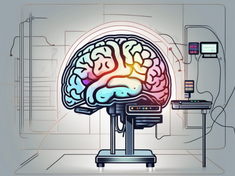
what do the lit up areas of fmri reveal
In the field of neuroscience, functional magnetic resonance imaging (fMRI) has revolutionized our understanding of the human brain. By analyzing the lit up areas in an fMRI scan, scientists can gain valuable insights into brain functioning and activity. In this article, we will delve into the fascinating world of fMRI and explore what these lit up areas reveal.
Understanding the Basics of fMRI
Before we dive into the intricacies of interpreting fMRI results, let’s start by understanding the fundamentals of this powerful imaging technique. fMRI is a non-invasive neuroimaging method that allows researchers to observe changes in blood flow and oxygen levels in the brain. By detecting these changes, fMRI can map brain activity and identify regions associated with specific cognitive processes or tasks.
Furthermore, fMRI has revolutionized the field of neuroscience by providing researchers with a window into the inner workings of the human brain. This technology has enabled scientists to study brain function in real-time, offering insights into various mental processes such as perception, memory, and decision-making.
The Science Behind fMRI
An fMRI machine uses a strong magnetic field and radio waves to generate detailed images of the brain. When certain brain areas become active, they require more oxygen and nutrients. To satisfy this increased demand, blood flow to those areas intensifies, resulting in a measurable change in the magnetic properties of the blood. By tracking these changes, fMRI can pinpoint regions of the brain that are engaged during a specific task or stimulus.
Moreover, fMRI technology has advanced our understanding of brain disorders and neurological conditions. By studying the brain activity patterns of individuals with conditions such as Alzheimer’s disease, schizophrenia, or depression, researchers can gain valuable insights into the underlying mechanisms of these disorders, potentially leading to improved diagnostic tools and treatment strategies.
Key Components of an fMRI Scan
An fMRI scan consists of several important components that work together to provide valuable data. Firstly, a powerful magnet produces the magnetic field required for imaging. Secondly, gradient coils enable precise spatial localization of brain activity. Finally, a radiofrequency coil detects radio waves emitted by the magnetized blood, which are then converted into detailed brain images by a computer.
Additionally, the data obtained from fMRI scans can be further analyzed using advanced computational techniques such as machine learning algorithms. These methods allow researchers to extract complex patterns of brain activity and connectivity, providing a deeper understanding of how different regions of the brain interact and communicate with each other during various cognitive tasks.
Interpreting the ‘Lit Up’ Areas in fMRI
Now that we have a foundation of understanding, let’s explore how to interpret the illuminated regions observed in an fMRI scan. It is important to note that these lit up areas do not represent an exact mapping of thoughts or emotions, but rather an indirect measurement of brain activity.
When looking at the colorful images produced by fMRI scans, it’s essential to understand that the brain is a highly intricate organ with a multitude of functions. The lit up areas, also known as activation clusters, indicate areas where there is a change in blood flow, suggesting increased neural activity. However, this activity is not always straightforward to interpret, as different regions of the brain can be involved in multiple cognitive processes simultaneously.
The Role of Blood Flow in fMRI
The lit up areas in an fMRI scan primarily reflect changes in blood flow. However, it is crucial to remember that increased blood flow does not necessarily equate to increased cognitive ability. The brain consists of a complex network of interconnected regions, and changes in blood flow might be a consequence of underlying processes that are not directly related to the task being performed.
Moreover, the relationship between blood flow and neural activity is not always linear. In some cases, a decrease in blood flow can also signify neural activity, a phenomenon known as the neurovascular coupling. This intricate dance between blood flow and neural firing is a constant area of study and refinement in the field of neuroimaging.
Decoding Brain Activity Through fMRI
Researchers use various analytical techniques to decode the patterns of brain activity revealed by fMRI. By employing advanced statistical methods, they can identify brain regions that consistently exhibit similar activation patterns across individuals, giving us valuable insights into how the brain is organized and functions.
One common method used in fMRI analysis is known as functional connectivity, which examines how different brain regions communicate with each other during specific tasks or at rest. This approach helps researchers map out the intricate networks that underlie various cognitive functions, shedding light on the complex interplay between different regions of the brain.
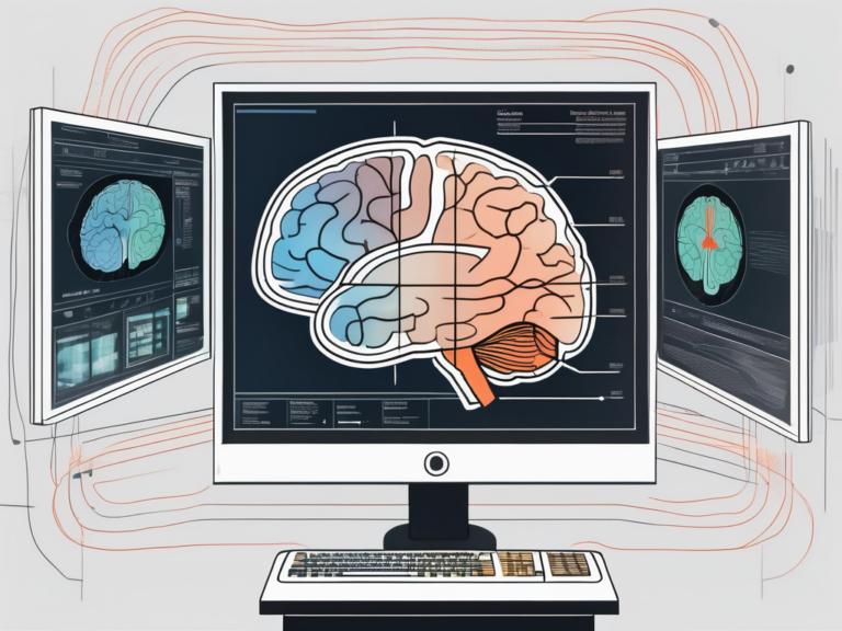
The Significance of Different Colors in fMRI
When viewing fMRI images, you may notice different colors associated with the various lit up areas. Understanding the significance of these colors can enhance our interpretation of fMRI results.
Functional Magnetic Resonance Imaging (fMRI) is a powerful tool that allows researchers to observe brain activity by measuring changes in blood flow. The colors in fMRI images play a crucial role in representing this data in a visually intuitive manner, aiding in the identification of active brain regions.
Color Coding in fMRI: What Does it Mean?
The colors in an fMRI image are assigned based on statistical values, indicating the strength of brain activity in different regions. Warm colors such as red, orange, and yellow represent higher activation, while cool colors like blue and green indicate lower levels of activation. These color representations allow researchers to visually identify and differentiate areas of interest in the brain.
Moreover, the color scale used in fMRI images is often standardized to ensure consistency across studies and research institutions. This standardization enables researchers to compare results effectively and facilitates the pooling of data for meta-analyses, contributing to the advancement of our understanding of the brain.
The Relationship Between Colors and Brain Regions
It is important to remember that colors in fMRI images do not directly correspond to specific brain functions or emotions. Instead, they indicate relative levels of activation in different brain regions. The interpretation of these colors is reliant on the context of the study and the experimental design.
Furthermore, advancements in fMRI technology have led to the development of sophisticated analytical tools that can extract detailed information from these color-coded images. Researchers can now perform complex analyses, such as functional connectivity studies, to investigate how different brain regions communicate with each other, providing insights into the underlying neural networks involved in various cognitive processes.
Limitations and Misconceptions About fMRI
While fMRI is a powerful tool, it is crucial to acknowledge its limitations and dispel common misconceptions surrounding its use.
Functional Magnetic Resonance Imaging (fMRI) has revolutionized the field of neuroscience by allowing researchers to non-invasively observe brain activity in real-time. By measuring changes in blood flow and oxygen levels, fMRI provides valuable insights into how different regions of the brain are involved in various cognitive tasks and behaviors.
Common Misunderstandings About fMRI Results
One common misconception is the belief that fMRI can read minds or accurately identify specific thoughts or intentions. It is essential to understand that fMRI results provide statistical associations between brain activity and specific tasks, rather than individual thoughts or intentions.
Furthermore, fMRI studies often involve group averages, meaning that individual variations in brain activity may not be fully captured. Factors such as participant variability, task design, and data analysis techniques can all influence the interpretation of fMRI results.
The Limitations of fMRI Technology
Another limitation of fMRI stems from its spatial and temporal resolution. While fMRI can produce detailed images of brain activity, it cannot capture activity at the level of individual neurons. Additionally, fMRI has a slight time delay in capturing changes in brain activity, limiting its ability to capture rapid cognitive processes.
Despite these limitations, fMRI remains a valuable tool for studying the brain and has contributed significantly to our understanding of cognition, emotion, and neurological disorders. Ongoing advancements in fMRI technology continue to enhance its capabilities, allowing researchers to delve deeper into the complexities of the human brain.
The Future of fMRI and Brain Imaging
As technology advances, so does our ability to delve deeper into the functioning of the human brain. Exciting developments in fMRI technology open the door to new possibilities and potential applications.
One of the most intriguing aspects of the future of fMRI and brain imaging lies in the realm of real-time imaging. Researchers are actively working on developing techniques that would allow for the monitoring of brain activity as it happens, providing a dynamic and detailed view of cognitive processes. This real-time fMRI could revolutionize fields such as neurofeedback training and brain-computer interfaces, offering unprecedented insights into brain function.
Recent Advances in fMRI Technology
Advancements in fMRI techniques, such as high-field strength magnets and multi-modal imaging approaches, allow for more detailed and precise mapping of brain activity. Furthermore, advanced analytical methods and machine learning algorithms are enhancing our ability to extract meaningful information from fMRI data.
Another cutting-edge development in fMRI technology is the integration of virtual reality (VR) environments. By immersing participants in realistic virtual scenarios while undergoing fMRI scans, researchers can study brain responses in highly ecologically valid settings. This fusion of VR technology with fMRI holds immense potential for studying complex behaviors and cognitive processes in a controlled yet naturalistic manner.
Potential Future Applications of fMRI
Looking forward, fMRI holds promise in various fields. It may contribute to improved understanding and treatment of neurological and psychiatric disorders, aid in the development of neurofeedback therapies, and provide insights into the effects of interventions such as medication or cognitive training on brain activity.
Furthermore, the combination of fMRI with other imaging modalities, such as electroencephalography (EEG) and magnetoencephalography (MEG), is opening up new avenues for multimodal brain imaging. By integrating data from different imaging techniques, researchers can overcome the limitations of each method individually and gain a more comprehensive understanding of brain function.
In conclusion, the lit up areas observed in an fMRI scan reveal valuable insights into brain functioning and activity. By understanding the basics of fMRI, interpreting the lit up regions, recognizing the significance of different colors, and acknowledging the limitations and future possibilities, we can harness the power of fMRI to deepen our understanding of the complex organ that is the human brain.