why is fmri better than mri
In the field of medical imaging, Magnetic Resonance Imaging (MRI) and Functional Magnetic Resonance Imaging (fMRI) are invaluable tools that revolutionized the diagnosis and understanding of various conditions and diseases. While both MRI and fMRI utilize powerful magnets and radio waves to create detailed images of the body’s internal structures, there are fundamental differences between the two techniques. This article aims to explore the intricacies of MRI and fMRI, highlighting the reasons why fMRI is considered superior in certain aspects.
Understanding the Basics of MRI and fMRI
MRI, or Magnetic Resonance Imaging, is a non-invasive imaging technique used widely in medicine to visualize the internal organs, tissues, and structures of the body. By employing a powerful magnetic field and radio waves, MRI scanners generate high-resolution, three-dimensional images that aid physicians in diagnosing and monitoring various conditions.
Fundamentally, MRI relies on the interaction of magnetic fields with hydrogen atoms present in the body. When the magnetic field is applied, these atoms align, and radio waves are used to perturb this alignment, causing them to emit signals. By analyzing these signals, detailed images of the body’s structures are generated.
In contrast, fMRI, or Functional Magnetic Resonance Imaging, builds upon the principles of MRI to observe brain activity in real-time. By measuring the changes in blood oxygenation levels in different regions of the brain, fMRI enables researchers and clinicians to assess brain functionality.
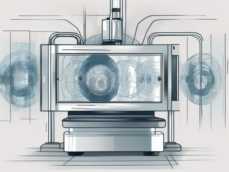
The Science Behind MRI
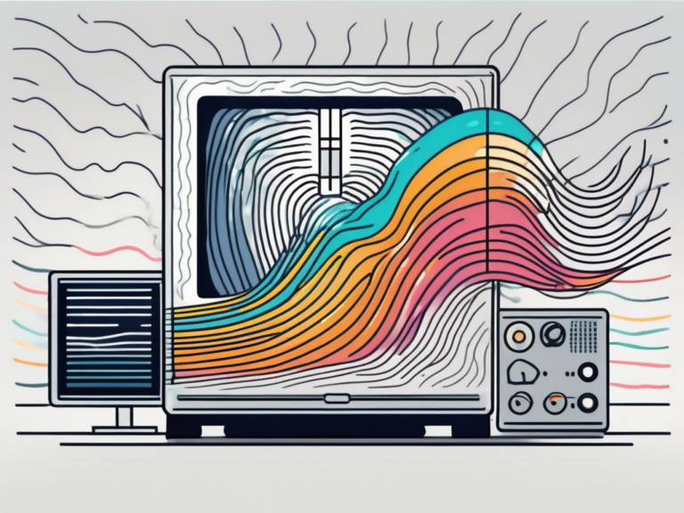
An MRI scanner consists of a large, cylindrical magnet that generates a strong, uniform magnetic field. Within this field, protons in the body align with the magnetic field. When radio frequency pulses are applied, protons absorb energy and then release it when the pulses cease. These released energy signals, combined with sophisticated algorithms, produce detailed images of the scanned body region.
But how does the MRI scanner differentiate between different types of tissues? Well, the hydrogen atoms in different tissues have slightly different magnetic properties. This variation allows the scanner to distinguish between, for example, muscle tissue and bone tissue. By manipulating the magnetic field and analyzing the signals emitted by the hydrogen atoms, the MRI scanner can create images with remarkable clarity and detail.
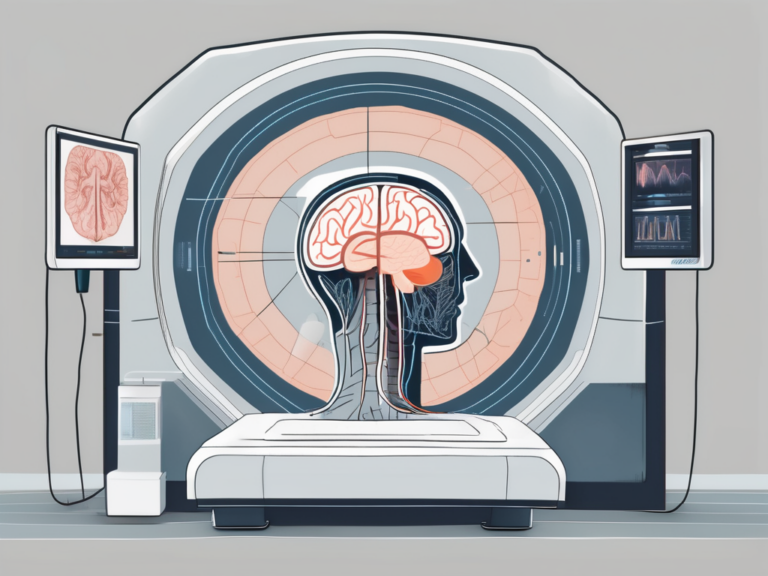
The Advanced Technology of fMRI
fMRI takes MRI a step further by utilizing the principle of blood oxygen level-dependent (BOLD) contrast. Changes in neural activity lead to increased blood flow to specific brain regions. This, in turn, affects the levels of oxygen in the blood. By detecting and measuring these variations in blood oxygenation, fMRI can identify the brain areas involved in certain cognitive processes or affected by neurological disorders.
One of the key challenges in fMRI is capturing images at an incredibly high speed. Since brain activity occurs within milliseconds, fMRI scanners need to acquire images rapidly to accurately capture the dynamic nature of brain function. This capability has been achieved through advancements in imaging technology, allowing researchers to track brain activation patterns in real-time.
Moreover, fMRI has revolutionized the field of neuroscience by enabling the mapping of complex neural networks. By studying brain activity during various tasks or stimuli, researchers can uncover the intricate connections between different brain regions and gain insights into how the brain processes information and controls behavior.
Key Differences Between MRI and fMRI
While both MRI and fMRI share similarities in terms of technology and imaging principles, there are significant differences that set them apart. Understanding these distinctions is crucial in appreciating the advantages of fMRI over traditional MRI.
Functional Versus Structural Imaging
One of the key differences between MRI and fMRI lies in the type of information they provide. MRI primarily focuses on providing detailed structural images of the body, enabling physicians to visualize anatomical abnormalities, tumors, or internal injuries.
In contrast, fMRI is uniquely suited for functional imaging by capturing the dynamic activity within the brain. It offers insights into how the brain processes information in real-time and helps elucidate the neural basis of various cognitive functions or disorders.
For example, fMRI can reveal which areas of the brain are activated when a person is engaged in a specific task, such as solving a math problem or listening to music. This information is invaluable in understanding the intricate workings of the human brain and can aid in the diagnosis and treatment of neurological conditions.
Time and Duration of Procedures
Another significant difference between MRI and fMRI lies in the time required for a complete scan. MRI scans typically take longer due to the need for high-resolution images and a comprehensive examination of the target area. The duration can range from 30 minutes to over an hour, limiting patient comfort and potentially affecting scan quality due to movement artifacts.
On the other hand, fMRI scans are relatively faster since they prioritize capturing real-time brain activation patterns. These scans usually last between 5 to 20 minutes, significantly reducing the discomfort and anxiety often associated with lengthy MRI procedures.
Moreover, the shorter duration of fMRI scans allows for multiple scans to be performed within a single session, providing researchers with a wealth of data to analyze and compare. This capability opens up new avenues for studying brain function and connectivity, leading to groundbreaking discoveries in the field of neuroscience.
Additionally, the reduced scan time of fMRI is particularly beneficial for pediatric patients or individuals who may have difficulty staying still for extended periods. By minimizing the time spent inside the scanner, fMRI offers a more comfortable and manageable experience for these individuals, ensuring accurate and reliable results.
Advantages of fMRI Over MRI
Given the differences outlined above, it becomes evident that fMRI offers significant advantages over traditional MRI, particularly in the realm of neuroscience and cognitive research.
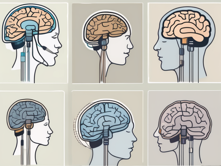
Enhanced Brain Mapping Capabilities
Due to its ability to track brain activation patterns, fMRI plays a pivotal role in mapping complex neurological processes. By correlating brain activities with specific stimuli or tasks, researchers can gain insights into how different brain regions communicate and orchestrate various cognitive functions.
This capability allows researchers to delve into the intricacies of brain disorders, such as Alzheimer’s disease, schizophrenia, or depression, leading to a deeper understanding of these conditions and potentially improved diagnosis and treatment strategies.
Moreover, the enhanced spatial resolution of fMRI enables researchers to pinpoint brain activity with remarkable precision. This detailed mapping is crucial for identifying subtle differences in brain function and connectivity, shedding light on the underlying mechanisms of various cognitive processes.
Non-Invasive Nature of fMRI
Additionally, fMRI operates on a non-invasive basis, making it a safer alternative to invasive procedures like positron emission tomography (PET) scans. By relying on magnetic fields and radio waves, fMRI avoids any exposure to potentially harmful ionizing radiation, reducing the associated risks involved in imaging procedures.
The non-invasive nature of fMRI also allows for repeated scans, enabling longitudinal studies and precise evaluations of treatment effectiveness over time.
Furthermore, the non-invasive nature of fMRI enhances its versatility in studying diverse populations, including children and individuals with certain medical conditions that may preclude them from undergoing invasive imaging techniques. This inclusivity broadens the scope of research possibilities and contributes to a more comprehensive understanding of brain function across different demographics.
Limitations of MRI in Comparison to fMRI
While MRI is an invaluable tool in medical imaging, it does have some limitations when compared to its functional counterpart, fMRI.
Despite its widespread use and effectiveness in capturing detailed anatomical images, MRI falls short in providing real-time functional data that can offer insights into how different parts of the body are actively responding to stimuli or tasks.
Lack of Functional Data in MRI
The primary drawback of MRI is its focus on anatomical structures rather than functional information. While MRI can provide detailed images of bodily organs and tissues, it cannot directly reveal how these structures are functioning or responding to specific stimuli.
The absence of functional data limits MRI’s utility in contexts where understanding real-time brain activity or assessing the impact of neurological interventions is crucial.
Conversely, fMRI, or functional magnetic resonance imaging, excels in capturing changes in blood flow and oxygen levels in the brain, offering a dynamic view of brain activity during various tasks or stimuli. This real-time functional data is crucial in fields such as neuroscience and cognitive psychology, where understanding brain function is paramount.
The Issue of Patient Comfort
MRI scans, typically longer in duration, can be challenging for patients, especially those with claustrophobia or anxiety disorders. The confined space inside the MRI machine, combined with the noise generated during the scan, can lead to discomfort and increased levels of stress.
fMRI, with its shorter scan durations, provides a significant advantage in terms of patient comfort. The reduced time spent inside the scanner helps alleviate anxiety, improving the overall experience for patients.
Furthermore, the noisy environment within the MRI machine can be unsettling for some patients, potentially affecting the quality of the images due to motion artifacts caused by involuntary movements. This issue is less pronounced in fMRI scans, where the shorter duration and quieter operation contribute to a more comfortable and motion artifact-free imaging experience.
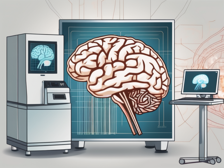
The Impact of fMRI on Medical Diagnosis and Research
The advent of functional Magnetic Resonance Imaging (fMRI) has significantly impacted medical diagnosis and research, offering new insights into the complexities of the human brain. This non-invasive imaging technique has revolutionized the field of neuroscience by allowing researchers and clinicians to observe brain activity in real-time.
fMRI works by measuring changes in blood flow and oxygenation levels in the brain, providing a detailed map of neural activity. This technology has become a cornerstone in the study of brain function, enabling professionals to better understand how different regions of the brain communicate and process information.
Improving Diagnostic Accuracy
fMRI’s ability to detect minute changes in brain activity enables clinicians to identify abnormalities, localize lesions, and improve diagnostic accuracy. By identifying areas of the brain affected by various conditions, fMRI assists in the diagnosis and treatment planning of neurological disorders, such as epilepsy or brain tumors. This enhances patient care and facilitates more targeted interventions.
Advancing Neurological Research
fMRI has ushered in a new era of neurological research by providing researchers with a powerful tool to study brain functions and disorders. By combining fMRI with cognitive tasks, scientists can investigate how the brain processes information, identify neural signatures of certain conditions, and uncover mechanisms underlying cognitive abilities.
This research has far-reaching implications, contributing to the development of more effective treatments and interventions for neurological and psychiatric disorders.
Furthermore, fMRI technology continues to evolve, with ongoing research focusing on improving spatial resolution and temporal accuracy. These advancements are crucial for enhancing the precision of fMRI scans and expanding their applications in both clinical and research settings. By pushing the boundaries of fMRI capabilities, scientists hope to unlock even deeper insights into the inner workings of the human brain.
The Future of fMRI and MRI
As technology continues to evolve, both fMRI and MRI are poised to benefit from numerous advancements, further widening their applications in healthcare.
Potential Developments in fMRI Technology
Future developments in fMRI technology aim to enhance the spatial and temporal resolution of the scans, allowing for more precise mapping of neural networks and real-time brain activity. This could potentially unlock a deeper understanding of how different brain regions interact and contribute to various cognitive processes and behaviors.
Researchers are also exploring the possibility of combining fMRI with other imaging techniques, such as electroencephalography (EEG) and magnetoencephalography (MEG), to create a more comprehensive picture of brain function. By integrating multiple modalities, scientists hope to overcome the limitations of each technique and gain a more holistic view of the brain’s complex dynamics.
The Ongoing Role of MRI in Healthcare
Despite the advancements in fMRI, MRI remains an indispensable tool in healthcare. Its ability to generate detailed anatomical images will continue to play a vital role in diagnosing and monitoring various medical conditions across multiple body systems.
However, the future of MRI holds exciting possibilities as well. Researchers are actively exploring the integration of artificial intelligence algorithms into MRI technology. By harnessing the power of machine learning, MRI scans can be analyzed more efficiently and accurately, leading to improved image quality and diagnostic precision. This could potentially revolutionize the field of radiology, enabling earlier detection and more personalized treatment plans for patients.
Moreover, advancements in MRI hardware and software are focused on enhancing patient comfort and reducing scan times. Innovations such as noise reduction technology and open MRI systems are making the scanning experience more pleasant for individuals, particularly those who may experience anxiety or claustrophobia.
In conclusion, both MRI and fMRI have revolutionized medical imaging and research. While MRI excels in providing high-resolution structural images, fMRI’s ability to observe brain functionality in real-time offers unrivaled advantages. With its enhanced brain mapping capabilities and non-invasive nature, fMRI has emerged as a powerful tool in understanding brain disorders and advancing neuroscience. Nonetheless, the ongoing advancements in both fMRI and MRI technology hold great promise for the future of medical imaging and research.