what does fmri stand for
fMRI, which stands for functional Magnetic Resonance Imaging, is a powerful tool used in the field of neuroscience. In this article, we will delve into the fascinating world of fMRI and explore its various applications, scientific principles, and future prospects. Let’s begin by understanding the acronym itself.
Understanding the Acronym: fMRI
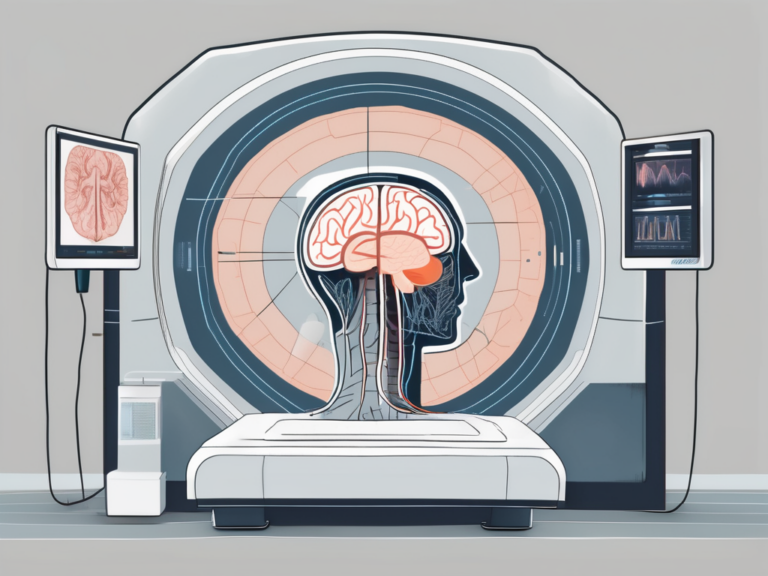
Before we dive into the depths of fMRI, let’s take a moment to decode the acronym itself. fMRI is short for functional Magnetic Resonance Imaging. The term “functional” indicates that this imaging technique allows us to observe the brain’s activity while it is performing specific functions. “Magnetic Resonance Imaging” refers to the method used to capture detailed images of the brain’s structure and function using powerful magnets and radio waves.
Functional Magnetic Resonance Imaging (fMRI) has become an indispensable tool in the field of neuroscience, offering researchers a non-invasive way to investigate brain function. This imaging technique measures changes in blood flow and oxygen levels in the brain, providing valuable insights into which areas of the brain are activated during different tasks or stimuli. By tracking these changes, scientists can create detailed maps of brain activity, helping to unravel the mysteries of cognition, emotion, and behavior.
The Full Form of fMRI
The full form of fMRI, as we have discussed, stands for functional Magnetic Resonance Imaging. This imaging technique has exponentially advanced our understanding of brain function and has revolutionized the field of neuroscience. By mapping neural activity with remarkable precision, fMRI has opened a window into the complex workings of the human brain.
Researchers and clinicians alike rely on fMRI to study various neurological and psychiatric conditions, such as Alzheimer’s disease, schizophrenia, and depression. By comparing brain activity patterns in healthy individuals to those with specific disorders, scientists can identify potential biomarkers and develop targeted treatments. The versatility and sensitivity of fMRI make it a powerful tool for both basic research and clinical applications, shaping our understanding of the brain’s inner workings.
The Origin of the Term fMRI
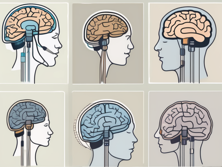
The term “fMRI” was first coined by Dr. Seiji Ogawa and his colleagues in the early 1990s. They discovered that changes in blood flow are closely linked to neural activity in the brain. By leveraging this relationship, Dr. Ogawa and his team devised a groundbreaking approach to map brain activity in real-time, leading to the birth of fMRI.
Dr. Ogawa’s pioneering work laid the foundation for a new era of brain imaging, allowing researchers to explore the dynamic interactions between brain regions and better understand the neural basis of cognition and behavior. The development of fMRI marked a significant milestone in neuroscience, offering a powerful tool to investigate the complexities of the human brain in health and disease.
The Science Behind fMRI
Now that we have a basic understanding of what fMRI stands for, let’s explore the scientific principles that underpin this fascinating imaging technique.
Functional Magnetic Resonance Imaging (fMRI) is a non-invasive neuroimaging technique that has revolutionized our ability to study the human brain in action. By measuring changes in blood flow and oxygenation levels, fMRI provides researchers with valuable insights into how different brain regions function during various cognitive tasks and emotional experiences.
Basic Principles of fMRI
Fundamentally, fMRI relies on a concept known as the blood-oxygen-level-dependent (BOLD) effect. When a specific brain region becomes active, there is an increase in blood flow to that region. This increased blood flow is accompanied by changes in the magnetic properties of the blood, especially the oxygenated and deoxygenated forms. By detecting these magnetic changes, fMRI can create images that reflect the brain’s activity levels.
Moreover, fMRI technology has advanced significantly over the years, allowing researchers to not only capture static snapshots of brain activity but also track dynamic changes in neural processes over time. This temporal dimension adds another layer of complexity to our understanding of how the brain responds to different stimuli and how neural networks interact to support various cognitive functions.
The Role of fMRI in Neuroscience
fMRI has emerged as a cornerstone technique in neuroscience research. It enables scientists to investigate various aspects of brain function, such as perception, memory, emotion, and decision-making. By examining the patterns of brain activity associated with specific tasks or mental states, fMRI helps unravel the intricate networks underlying human cognition and behavior.
Furthermore, the versatility of fMRI extends beyond basic research, with applications in clinical settings for diagnosing neurological disorders, monitoring treatment outcomes, and even guiding surgical interventions in delicate brain areas. The ability of fMRI to provide real-time feedback on brain activity has opened up new possibilities for personalized medicine and targeted therapies tailored to individual patients’ neural profiles.
Applications of fMRI
Having established a solid foundation in the science behind fMRI, let’s now explore its practical applications in different fields.
Functional Magnetic Resonance Imaging (fMRI) has revolutionized the way we understand the human brain and its functions. By measuring changes in blood flow, fMRI allows researchers to map brain activity in real-time, providing valuable insights into cognitive processes.
fMRI in Clinical Diagnosis
In the field of medicine, fMRI plays a crucial role in diagnosing and understanding various neurologic and psychiatric disorders. By observing the brain’s responses to certain stimuli, medical professionals can identify abnormalities and tailor treatments accordingly. fMRI has proven particularly valuable in assessing conditions like stroke, Alzheimer’s disease, schizophrenia, and depression.
Furthermore, fMRI is instrumental in presurgical planning, helping surgeons identify critical brain regions to avoid during procedures. This technology has significantly improved the success rates of delicate neurosurgical interventions, minimizing the risk of postoperative complications.
fMRI in Psychological Research
Psychological researchers have also embraced fMRI as a valuable tool. By examining brain activity, they can shed light on topics such as learning, memory, attention, and emotion. fMRI provides researchers with a unique opportunity to investigate the neural mechanisms underlying psychological processes, unraveling the mysteries of the human mind.
Moreover, fMRI studies have contributed to our understanding of developmental disorders such as autism spectrum disorder (ASD) and attention-deficit/hyperactivity disorder (ADHD). By comparing brain activity patterns in individuals with these conditions to neurotypical individuals, researchers can pinpoint areas of divergence, paving the way for targeted interventions and therapies.
The Process of an fMRI Scan
Now that we have explored the applications of fMRI, let’s turn our attention to the actual process of undergoing an fMRI scan.
Functional Magnetic Resonance Imaging (fMRI) is a powerful tool used in neuroscience to measure brain activity by detecting changes in blood flow. This non-invasive technique provides valuable insights into how different regions of the brain function and communicate with each other.
Preparing for an fMRI Scan
Prior to an fMRI scan, it is important to follow the instructions provided by the medical professionals conducting the procedure. Patients are typically required to remove any metal objects, such as jewelry or clothing with metallic components, as these can interfere with the magnetic field. Additionally, individuals with claustrophobia or other concerns may receive appropriate guidance or accommodations to ensure their comfort during the procedure.
Furthermore, it is crucial for patients to remain still during the scan to obtain clear and accurate images. Any movement can distort the results, making it essential for individuals to understand the importance of staying as motionless as possible throughout the procedure.
What Happens During an fMRI Scan
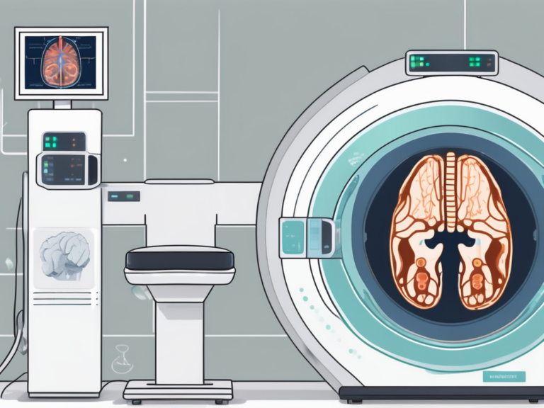
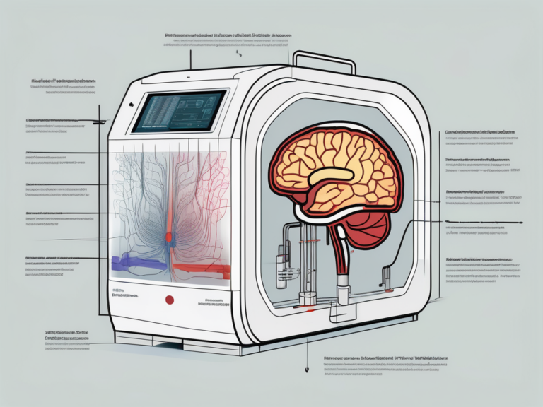
During an fMRI scan, the patient lies down on a comfortable bed that will move into the scanner. The most common type of fMRI is called BOLD fMRI, in which changes in blood oxygenation are detected. The patient is often asked to perform specific tasks or think about certain things while inside the scanner. Meanwhile, sophisticated sensors capture the changes in brain activity, allowing the creation of detailed images.
As the patient engages in different activities or cognitive tasks, the fMRI machine tracks the blood flow to various parts of the brain, highlighting areas that are more active during specific functions. This data is then processed using complex algorithms to generate colorful maps that represent brain activity patterns, providing researchers and healthcare professionals with valuable information for diagnosis and treatment planning.
Interpreting fMRI Results
Once the fMRI scan is completed, a skilled team of experts carefully analyzes the data and interprets the results. This process involves examining the intricate patterns of brain activity captured during the scan to draw meaningful conclusions about cognitive processes, emotions, and behaviors.
Furthermore, the interpretation of fMRI results often involves comparing the data with existing knowledge of brain function and structures. By referencing established neuroanatomical and neurophysiological principles, researchers can better understand the implications of the observed activation patterns and their relevance to specific mental functions.
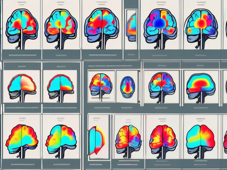
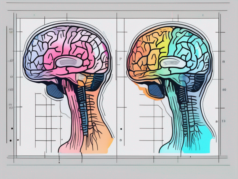
Understanding fMRI Images
fMRI images show different brain regions highlighted in certain colors, indicating their level of activation during the performed tasks or conditions. These images provide invaluable insights into the functional organization of the brain and can help identify any abnormalities or deviations from normal patterns of activity. Researchers can overlay these activation maps onto structural MRI images to precisely locate the activated regions within the brain, allowing for a more comprehensive analysis of the neural circuits involved in various cognitive processes.
Limitations and Challenges in fMRI Analysis
While fMRI has revolutionized neuroscience, it is important to acknowledge its limitations. The interpretation of fMRI results is intricately linked to the experimental design and statistical modeling employed. As with any scientific technique, rigorous methodology and critical analysis of results are essential to ensure accurate and meaningful interpretations. Researchers must also consider factors such as motion artifacts, physiological noise, and individual variability in brain structure and function, which can introduce complexities and confounds into the analysis.
Moreover, the field of fMRI research continually evolves, with ongoing debates and advancements in data processing techniques and analytical approaches. Staying abreast of these developments is crucial for researchers to enhance the reliability and validity of their findings. Collaborations between experts in neuroscience, psychology, and data science play a vital role in addressing the multifaceted challenges associated with fMRI analysis and interpretation, ultimately pushing the boundaries of our understanding of the human brain.
The Future of fMRI
As technology continues to advance, we can expect exciting developments in the field of fMRI.
Looking ahead, the future of fMRI holds great promise with the potential for groundbreaking advancements that could revolutionize our understanding of the brain. Researchers are exploring innovative ways to enhance the capabilities of fMRI technology, pushing the boundaries of what is currently possible.
Technological Advances in fMRI
Ongoing research efforts aim to refine fMRI technology and its applications. One such area of focus is increasing the spatial and temporal resolution of fMRI scans, which would provide even greater insight into the brain’s dynamics. The quest for higher resolution imaging is driven by the desire to capture the brain’s activity with unprecedented detail, allowing for more precise localization of neural processes.
Furthermore, the integration of artificial intelligence and machine learning algorithms into fMRI data analysis is a burgeoning field that shows immense potential. These advanced computational techniques have the capacity to extract deeper insights from fMRI data, potentially uncovering patterns and relationships that were previously undetectable.
Ethical Considerations in fMRI Use
As with any powerful tool, the use of fMRI presents ethical considerations that must be carefully addressed. Safeguarding privacy and confidentiality, ensuring informed consent, and weighing the potential risks and benefits are all crucial aspects of the responsible utilization of fMRI technology.
Moreover, the ethical implications of using fMRI in vulnerable populations, such as children or individuals with cognitive impairments, require special attention. Researchers and clinicians must navigate complex ethical dilemmas to ensure that the benefits of fMRI research outweigh any potential risks or harms to participants.
In conclusion, fMRI, or functional Magnetic Resonance Imaging, stands at the frontier of modern neuroscience. Through its ability to map brain activity, fMRI has allowed researchers to better understand the intricacies of the human mind. With ongoing technological advancements and ethical considerations, fMRI holds immense potential to further illuminate the mysteries of the brain and advance both scientific knowledge and patient care.