what is a drawback of having an fmri scan?
In recent years, functional magnetic resonance imaging (fMRI) has emerged as a valuable tool for understanding the complex workings of the human brain. By measuring changes in blood flow, fMRI allows researchers to identify brain regions that are active during different cognitive tasks or in response to various stimuli. This non-invasive technique has revolutionized neuroscience and provided invaluable insights into the inner workings of the mind. However, like any medical procedure, fMRI scans come with their own set of risks and drawbacks. In this article, we will explore the potential drawbacks of having an fMRI scan and discuss alternative options that individuals may consider.
Understanding the Basics of an fMRI Scan
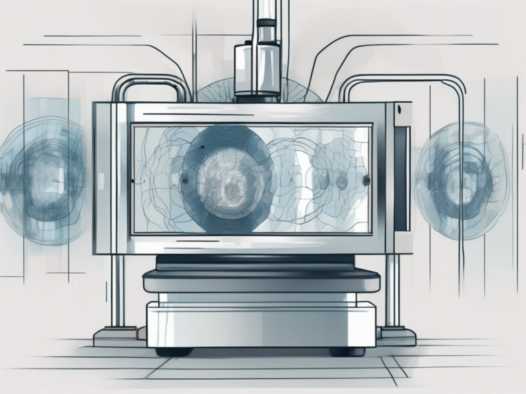
Before diving into the drawbacks, let’s take a moment to understand the basics of an fMRI scan. During the scan, the patient lies still inside a large cylindrical machine while a series of images are taken of their brain. These images, known as fMRI images, show the areas of the brain that are activated during specific tasks or conditions.
The Science Behind fMRI Scans
An fMRI scan relies on the principle that changes in neural activity are accompanied by changes in blood flow. When a particular brain region becomes active, blood flow to that area increases, bringing oxygen and nutrients needed for the increased metabolic demand. This change in blood flow can be detected by the fMRI scanner, allowing researchers to create detailed maps of the brain’s activity.
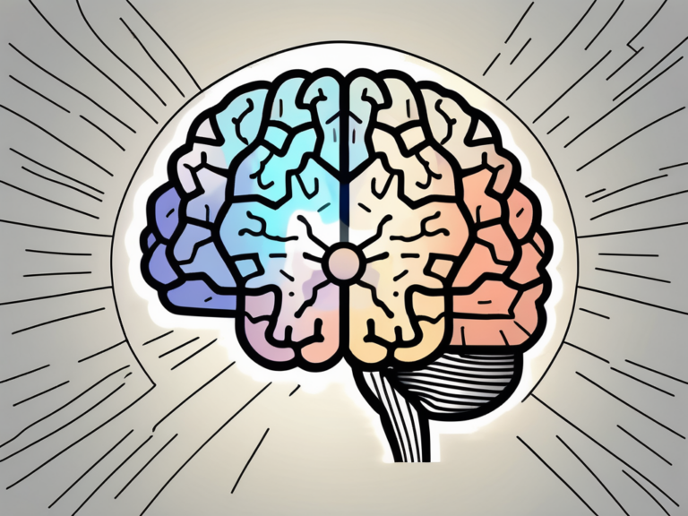
Furthermore, fMRI technology has advanced significantly over the years, allowing for higher resolution images and more precise mapping of brain activity. Researchers can now track not only which areas of the brain are activated during a task but also the sequence in which they are activated, providing a deeper understanding of the brain’s intricate processes.
Common Uses of fMRI Scans
fMRI scans have become invaluable tools in the field of neuroscience. They are widely used to study brain functions, such as language processing, memory, attention, emotion, and decision-making. fMRI scans have also been used to investigate neurological disorders, such as Alzheimer’s disease, Parkinson’s disease, and schizophrenia, providing valuable insights into the underlying mechanisms of these conditions.
Moreover, fMRI scans are not limited to studying brain function in healthy individuals or those with neurological disorders. They are also used in research on various psychological conditions, such as addiction, depression, and anxiety disorders. By observing the patterns of brain activity associated with these conditions, researchers can develop more effective treatments and interventions to improve the lives of affected individuals.
Potential Risks and Drawbacks of fMRI Scans
While fMRI scans are generally considered safe, there are some potential drawbacks and risks associated with this procedure. It is important to be aware of these factors before deciding to undergo an fMRI scan.
One potential drawback of fMRI scans is the physical discomfort and claustrophobia that some individuals may experience. The fMRI scanner is a large and noisy machine, which can cause discomfort and anxiety for certain patients. The confined space inside the machine can also trigger claustrophobia, making it difficult for some individuals to tolerate the procedure. It is important to inform the healthcare provider if you have any concerns or previous experiences with claustrophobia, as they may be able to provide strategies to help alleviate these discomforts.
Another limitation of fMRI scans is the challenge in accurately interpreting the images. While the technology has advanced significantly, interpreting fMRI images still requires expertise and knowledge in the field of neuroscience. False positives and false negatives can occur, leading to incorrect conclusions about brain activity. Therefore, it is crucial to have trained professionals evaluate the results to avoid misinterpretation. This ensures that the findings are accurately understood and can be used to inform further research or medical decisions.
Aside from physical discomfort and interpretation challenges, the high cost of fMRI scans can also be a significant drawback for some individuals. These scans can be quite expensive, making them less accessible for individuals with limited financial resources or without adequate healthcare coverage. The cost includes not only the equipment and technical expertise required for the procedure but also the professional interpretation of the results. This financial burden can limit the access to this technology, potentially preventing individuals from benefiting from its diagnostic or research potential.
It is important to weigh these potential risks and drawbacks against the potential benefits of undergoing an fMRI scan. Discussing these factors with your healthcare provider can help you make an informed decision that takes into account your individual circumstances and priorities.
Comparing fMRI with Other Brain Imaging Techniques
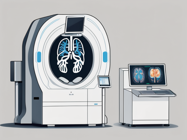
It is important to consider fMRI scans in the broader context of other brain imaging techniques. Let’s compare fMRI with two commonly used imaging modalities: CT scans and PET scans.
When delving into the realm of brain imaging techniques, it becomes evident that each method offers unique insights into the intricate workings of the human brain. While fMRI excels in capturing real-time changes in blood flow associated with neural activity, it is essential to explore how other imaging modalities complement and enhance our understanding of brain structure and function.
fMRI vs. CT Scans
Unlike fMRI, which measures changes in blood flow, CT (computed tomography) scans use a series of X-ray images to create detailed cross-sectional images of the brain. CT scans are valuable tools for detecting structural abnormalities such as tumors, hemorrhages, or bone fractures within the brain. The ability of CT scans to provide precise anatomical details makes them indispensable in emergency settings where swift diagnosis is crucial. However, it is important to note that CT scans do not offer insights into brain function or dynamic processes like fMRI scans do.
fMRI vs. PET Scans
Similar to fMRI, PET (positron emission tomography) scans also capture brain activity, albeit through a different mechanism. While fMRI tracks changes in blood flow, PET scans involve the injection of a radioactive substance that allows visualization of metabolic processes in the brain. This metabolic information provided by PET scans is particularly valuable in studying conditions such as epilepsy, Alzheimer’s disease, and certain psychiatric disorders. Despite the wealth of data they offer, PET scans come with the drawback of exposing patients to radiation, necessitating careful consideration of the risks and benefits associated with this imaging modality.
Addressing the Drawbacks: Improvements and Alternatives
Despite the drawbacks mentioned earlier, there have been notable improvements in fMRI technology and alternative brain imaging techniques. It is crucial to stay informed about the latest advancements in the field of neuroimaging to make informed decisions about diagnostic and research tools.
One significant area of improvement in fMRI technology is the development of higher field strength scanners. These scanners offer increased signal-to-noise ratio, resulting in clearer images and more precise data. Additionally, the integration of real-time fMRI technology allows for immediate feedback during brain scans, enabling researchers to adjust parameters for better accuracy.
Technological Advancements in fMRI Scans
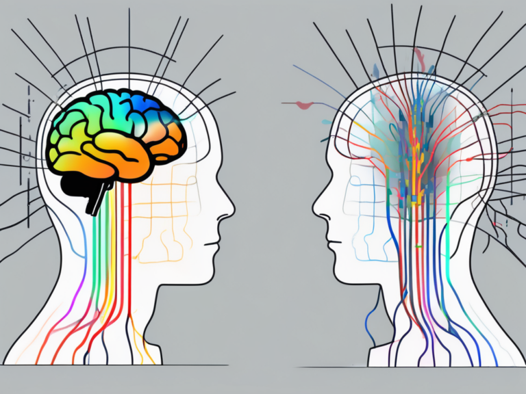
Researchers are continuously working on improving fMRI technology to enhance image quality, reduce noise, and improve the accuracy of interpretation. Advances in machine learning and data analysis techniques are also helping researchers unlock more information from fMRI scans, increasing their diagnostic value. The use of multi-modal imaging, combining fMRI with other imaging modalities such as structural MRI or diffusion tensor imaging, provides a more comprehensive understanding of brain structure and function.
Furthermore, the development of portable fMRI systems is revolutionizing the field by allowing for brain imaging in naturalistic settings, such as during real-world tasks or in populations where traditional fMRI scanners are not feasible. These advancements open up new possibilities for studying brain function in diverse populations and environments.
Exploring Alternative Brain Imaging Techniques
There are several alternative brain imaging techniques that individuals can consider based on their specific needs or concerns. These may include techniques such as electroencephalography (EEG), magnetoencephalography (MEG), or functional near-infrared spectroscopy (fNIRS). Each of these techniques has its own advantages and limitations, and it is important to consult with a healthcare professional to determine which method is best suited for individual circumstances.
EEG, for example, offers excellent temporal resolution, making it ideal for studying rapid brain processes such as event-related potentials. MEG, on the other hand, provides superior spatial resolution for localizing brain activity with millisecond precision. fNIRS is a non-invasive technique that measures changes in blood oxygen levels in the brain, offering portability and ease of use for various applications.
Making an Informed Decision: Is fMRI Right for You?
Before undergoing an fMRI scan or considering alternative brain imaging techniques, it is essential to carefully evaluate the potential benefits and drawbacks.
Factors to Consider Before Undergoing an fMRI Scan
Factors to consider include the specific medical condition or research question, available healthcare coverage, individual tolerance for claustrophobic environments, and individual preferences for risk, cost, and convenience. It is crucial to have an open and honest discussion with a healthcare provider who can provide personalized advice based on the individual’s unique circumstances.
Discussing Your Options with Your Healthcare Provider
Ultimately, the decision of whether to undergo an fMRI scan or opt for an alternative brain imaging technique rests with the individual and their healthcare provider. It is essential to engage in a meaningful conversation with the healthcare provider, asking questions, expressing concerns, and fully understanding the potential benefits and drawbacks of each option. With expert guidance, individuals can make informed decisions that align with their healthcare needs and personal preferences.
When considering the potential benefits of an fMRI scan, it is important to note that this imaging technique offers a non-invasive way to examine the brain’s structure and function. By providing detailed images of brain activity, fMRI scans have revolutionized the field of neuroscience, allowing researchers and healthcare professionals to gain valuable insights into various neurological conditions and cognitive processes.
However, it is equally important to acknowledge the limitations and drawbacks of fMRI scans. For instance, the cost of an fMRI scan can be a significant factor to consider, especially if healthcare coverage is limited. Additionally, some individuals may experience claustrophobia or discomfort in the confined space of the MRI machine, making it necessary to explore alternative imaging techniques that can provide similar insights without causing distress.
Moreover, it is crucial to recognize that fMRI scans are not without their limitations. While they can provide detailed images of brain activity, they do not provide a direct measurement of neural activity. Instead, fMRI scans rely on changes in blood flow and oxygenation levels to infer brain activity. This indirect measurement can introduce some degree of uncertainty and may not capture the full complexity of neural processes.
Therefore, when making a decision about whether to undergo an fMRI scan, it is vital to carefully weigh the potential benefits and drawbacks, considering individual circumstances, preferences, and healthcare needs. By engaging in informed decision-making and discussing options with a healthcare provider, individuals can navigate the complexities of brain imaging techniques, ensuring that they make choices that align with their unique situations and contribute to the advancement of neuroscience.